2FGV

X-ray crystal structure of HIV-1 Protease T80N variant in complex with the inhibitor saquinavir used to explore the role of invariant Thr80 in HIV-1 protease structure, function, and viral infectivity.


Experimental Data Snapshot

  • Method: X-RAY DIFFRACTION
  • Resolution: 1.50 Å
  • R-Value Free: 0.197 
  • R-Value Work: 0.171 
  • R-Value Observed: 0.173 

wwPDB Validation   3D Report Full Report


This is version 1.6 of the entry. See complete history


Literature

Role of invariant Thr80 in human immunodeficiency virus type 1 protease structure, function, and viral infectivity.

Foulkes, J.E.Prabu-Jeyabalan, M.Cooper, D.Henderson, G.J.Harris, J.Swanstrom, R.Schiffer, C.A.

(2006) J Virol 80: 6906-6916

  • DOI: https://doi.org/10.1128/JVI.01900-05
  • Primary Citation of Related Structures:  
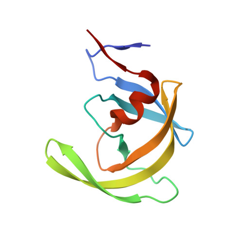
    2FGU, 2FGV

  • PubMed Abstract: 

    Sequence variability associated with human immunodeficiency virus type 1 (HIV-1) is useful for inferring structural and/or functional constraints at specific residues within the viral protease. Positions that are invariant even in the presence of drug selection define critically important residues for protease function. While the importance of conserved active-site residues is easily understood, the role of other invariant residues is not. This work focuses on invariant Thr80 at the apex of the P1 loop of HIV-1, HIV-2, and simian immunodeficiency virus protease. In a previous study, we postulated, on the basis of a molecular dynamics simulation of the unliganded protease, that Thr80 may play a role in the mobility of the flaps of protease. In the present study, both experimental and computational methods were used to study the role of Thr80 in HIV protease. Three protease variants (T80V, T80N, and T80S) were examined for changes in structure, dynamics, enzymatic activity, affinity for protease inhibitors, and viral infectivity. While all three variants were structurally similar to the wild type, only T80S was functionally similar. Both T80V and T80N had decreased the affinity for saquinavir. T80V significantly decreased the ability of the enzyme to cleave a peptide substrate but maintained infectivity, while T80N abolished both activity and viral infectivity. Additionally, T80N decreased the conformational flexibility of the flap region, as observed by simulations of molecular dynamics. Taken together, these data indicate that HIV-1 protease functions best when residue 80 is a small polar residue and that mutations to other amino acids significantly impair enzyme function, possibly by affecting the flexibility of the flap domain.


  • Organizational Affiliation

    Department of Biochemistry and Molecular Pharmacology, University of Massachusetts Medical School, 364 Plantation Street, Worcester, 01605, USA.


Macromolecules
Find similar proteins by:  (by identity cutoff)  |  3D Structure
Entity ID: 1
MoleculeChains Sequence LengthOrganismDetailsImage
Protease
A, B
99Human immunodeficiency virus 1Mutation(s): 3 
Gene Names: GAG
EC: 3.4.23.16
UniProt
Find proteins for P03369 (Human immunodeficiency virus type 1 group M subtype B (isolate ARV2/SF2))
Explore P03369 
Go to UniProtKB:  P03369
Entity Groups  
Sequence Clusters30% Identity50% Identity70% Identity90% Identity95% Identity100% Identity
UniProt GroupP03369
Sequence Annotations
Expand
  • Reference Sequence
Small Molecules
Ligands 6 Unique
IDChains Name / Formula / InChI Key2D Diagram3D Interactions
DIQ
Query on DIQ

Download Ideal Coordinates CCD File 
J [auth A]2-METHYL-DECAHYDRO-ISOQUINOLINE-3-CARBOXYLIC ACID
C11 H19 N O2
YKQLYDHGCIEUMU-AEJSXWLSSA-N
QNC
Query on QNC

Download Ideal Coordinates CCD File 
G [auth A]quinoline-2-carboxylic acid
C10 H7 N O2
LOAUVZALPPNFOQ-UHFFFAOYSA-N
HPH
Query on HPH

Download Ideal Coordinates CCD File 
I [auth A](2S)-2-amino-3-phenylpropane-1,1-diol
C9 H13 N O2
IFTWVTAUEXLCHB-QMMMGPOBSA-N
ASN
Query on ASN

Download Ideal Coordinates CCD File 
H [auth A]ASPARAGINE
C4 H8 N2 O3
DCXYFEDJOCDNAF-REOHCLBHSA-N
PO4
Query on PO4

Download Ideal Coordinates CCD File 
C [auth A]
D [auth A]
E [auth A]
F [auth A]
L [auth B]
C [auth A],
D [auth A],
E [auth A],
F [auth A],
L [auth B],
M [auth B],
N [auth B]
PHOSPHATE ION
O4 P
NBIIXXVUZAFLBC-UHFFFAOYSA-K
NTB
Query on NTB

Download Ideal Coordinates CCD File 
K [auth A]TERTIARY-BUTYLAMINE
C4 H11 N
YBRBMKDOPFTVDT-UHFFFAOYSA-N
Experimental Data & Validation

Experimental Data

  • Method: X-RAY DIFFRACTION
  • Resolution: 1.50 Å
  • R-Value Free: 0.197 
  • R-Value Work: 0.171 
  • R-Value Observed: 0.173 
  • Space Group: P 21 21 21
Unit Cell:
Length ( Å )Angle ( ˚ )
a = 50.52α = 90
b = 58.18β = 90
c = 61.52γ = 90
Software Package:
Software NamePurpose
REFMACrefinement
SCALEPACKdata scaling
AMoREphasing

Structure Validation

View Full Validation Report


Entry History 

Deposition Data

Revision History  (Full details and data files)

  • Version 1.0: 2006-11-07
    Type: Initial release
  • Version 1.1: 2008-05-01
    Changes: Version format compliance
  • Version 1.2: 2011-07-13
    Changes: Non-polymer description, Version format compliance
  • Version 1.3: 2017-10-18
    Changes: Refinement description
  • Version 1.4: 2018-01-24
    Changes: Structure summary
  • Version 1.5: 2021-10-20
    Changes: Database references, Derived calculations
  • Version 1.6: 2023-08-30
    Changes: Data collection, Refinement description