2FGE

Crystal structure of presequence protease PreP from Arabidopsis thaliana


Experimental Data Snapshot

  • Method: X-RAY DIFFRACTION
  • Resolution: 2.10 Å
  • R-Value Free: 0.256 
  • R-Value Work: 0.206 
  • R-Value Observed: 0.209 

wwPDB Validation   3D Report Full Report


This is version 1.3 of the entry. See complete history


Literature

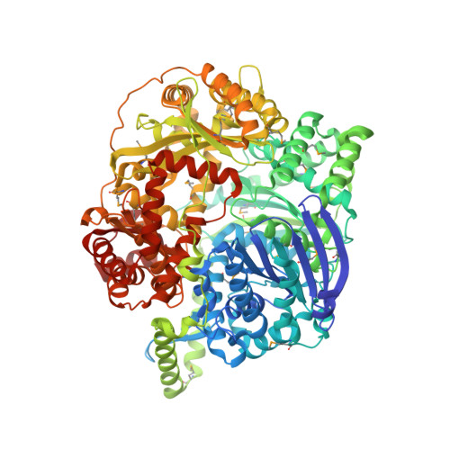
The closed structure of presequence protease PreP forms a unique 10 000 A(3) chamber for proteolysis

Johnson, K.A.Bhushan, S.Hallberg, B.M.Frohn, A.Glaser, E.Eneqvist, T.

(2006) EMBO J 25: 1977-1986

  • DOI: https://doi.org/10.1038/sj.emboj.7601080
  • Primary Citation of Related Structures:  
    2FGE

  • PubMed Abstract: 

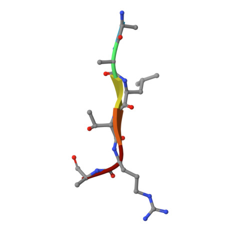
    Presequence protease PreP is a novel protease that degrades targeting peptides as well as other unstructured peptides in both mitochondria and chloroplasts. The first structure of PreP from Arabidopsis thaliana refined at 2.1 Angstroms resolution shows how the 995-residue polypeptide forms a unique proteolytic chamber of more than 10,000 Angstroms(3) in which the active site resides. Although there is no visible opening to the chamber, a peptide is bound to the active site. The closed conformation places previously unidentified residues from the C-terminal domain at the active site, separated by almost 800 residues in sequence to active site residues located in the N-terminal domain. Based on the structure, a novel mechanism for proteolysis is proposed involving hinge-bending motions that cause the protease to open and close in response to substrate binding. In support of this model, cysteine double mutants designed to keep the chamber covalently locked show no activity under oxidizing conditions. The manner in which substrates are processed inside the chamber is reminiscent of the proteasome; therefore, we refer to this protein as a peptidasome.


  • Organizational Affiliation

    Department of Biochemistry and Biophysics, Arrhenius Laboratories for Natural Sciences, Stockholm University, Sweden.


Macromolecules
Find similar proteins by:  (by identity cutoff)  |  3D Structure
Entity ID: 1
MoleculeChains Sequence LengthOrganismDetailsImage
zinc metalloprotease (insulinase family)
A, B
995Arabidopsis thalianaMutation(s): 21 
UniProt
Find proteins for Q9LJL3 (Arabidopsis thaliana)
Explore Q9LJL3 
Go to UniProtKB:  Q9LJL3
Entity Groups  
Sequence Clusters30% Identity50% Identity70% Identity90% Identity95% Identity100% Identity
UniProt GroupQ9LJL3
Sequence Annotations
Expand
  • Reference Sequence

Find similar proteins by:  Sequence   |   3D Structure  

Entity ID: 2
MoleculeChains Sequence LengthOrganismDetailsImage
nonspecific peptide AALTRAC [auth D],
D [auth E]
6N/AMutation(s): 0 
Sequence Annotations
Expand
  • Reference Sequence
Small Molecules
Modified Residues  1 Unique
IDChains TypeFormula2D DiagramParent
MSE
Query on MSE
A, B
L-PEPTIDE LINKINGC5 H11 N O2 SeMET
Experimental Data & Validation

Experimental Data

  • Method: X-RAY DIFFRACTION
  • Resolution: 2.10 Å
  • R-Value Free: 0.256 
  • R-Value Work: 0.206 
  • R-Value Observed: 0.209 
  • Space Group: P 21 21 21
Unit Cell:
Length ( Å )Angle ( ˚ )
a = 110.834α = 90
b = 114.332β = 90
c = 162.984γ = 90
Software Package:
Software NamePurpose
DENZOdata reduction
SCALEPACKdata scaling
SHELXSphasing
REFMACrefinement

Structure Validation

View Full Validation Report


Entry History 

Deposition Data

Revision History  (Full details and data files)

  • Version 1.0: 2006-05-16
    Type: Initial release
  • Version 1.1: 2008-05-01
    Changes: Version format compliance
  • Version 1.2: 2011-07-13
    Changes: Version format compliance
  • Version 1.3: 2021-10-20
    Changes: Database references, Derived calculations