2FDP

Crystal structure of beta-secretase complexed with an amino-ethylene inhibitor


Experimental Data Snapshot

  • Method: X-RAY DIFFRACTION
  • Resolution: 2.50 Å
  • R-Value Free: 0.254 
  • R-Value Work: 0.225 
  • R-Value Observed: 0.227 

wwPDB Validation   3D Report Full Report


Ligand Structure Quality Assessment 


This is version 1.2 of the entry. See complete history


Literature

Aminoethylenes: a tetrahedral intermediate isostere yielding potent inhibitors of the aspartyl protease BACE-1.

Yang, W.Lu, W.Lu, Y.Zhong, M.Sun, J.Thomas, A.E.Wilkinson, J.M.Fucini, R.V.Lam, M.Randal, M.Shi, X.P.Jacobs, J.W.McDowell, R.S.Gordon, E.M.Ballinger, M.D.

(2006) J Med Chem 49: 839-842

  • DOI: https://doi.org/10.1021/jm0509142
  • Primary Citation of Related Structures:  
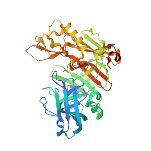
    2FDP

  • PubMed Abstract: 

    A series of novel beta-site amyloid precursor protein cleaving enzyme (BACE-1) inhibitors containing an aminoethylene (AE) tetrahedral intermediate isostere were synthesized and evaluated in comparison to corresponding hydroxyethylene (HE) compounds. Enzymatic inhibitory values were similar for both isosteres, as were structure-activity relationships with respect to stereochemical preference and substituent variation (P2/P3, P1, and P2'); however, the AE compounds were markedly more potent in a cell-based assay for reduction of beta-secretase activity. The incorporation of preferred P2/P3, P1, and P2' substituents into the AE pharmacophore yielded compound 7, which possessed enzymatic and cell assay IC(50)s of 26 nM and 180 nM, respectively. A three-dimensional crystal structure of 7 in complex with BACE-1 revealed that the amino group of the inhibitor core engages the catalytic aspartates in a manner analogous to hydroxyl groups in HE inhibitors. The AE isostere class represents a promising advance in the development of BACE-1 inhibitors.


  • Organizational Affiliation

    Departments of Chemistry, Biochemistry, and Structural Biology, Sunesis Pharmaceuticals Incorporated, 341 Oyster Point Boulevard, South San Francisco, CA 94080, USA. wenjin@sunesis.com


Macromolecules
Find similar proteins by:  (by identity cutoff)  |  3D Structure
Entity ID: 1
MoleculeChains Sequence LengthOrganismDetailsImage
Beta-secretase 1
A, B, C
388Homo sapiensMutation(s): 0 
Gene Names: BACE1
EC: 3.4.23.46
UniProt & NIH Common Fund Data Resources
Find proteins for P56817 (Homo sapiens)
Explore P56817 
Go to UniProtKB:  P56817
PHAROS:  P56817
GTEx:  ENSG00000186318 
Entity Groups  
Sequence Clusters30% Identity50% Identity70% Identity90% Identity95% Identity100% Identity
UniProt GroupP56817
Sequence Annotations
Expand
  • Reference Sequence
Small Molecules
Ligands 1 Unique
IDChains Name / Formula / InChI Key2D Diagram3D Interactions
FRP
Query on FRP

Download Ideal Coordinates CCD File 
D [auth A],
E [auth B],
F [auth C]
N1-((2S,3S,5R)-3-AMINO-6-(4-FLUOROPHENYLAMINO)-5-METHYL-6-OXO-1-PHENYLHEXAN-2-YL)-N3,N3-DIPROPYLISOPHTHALAMIDE
C33 H41 F N4 O3
PZHFCAIXAVDQAK-STQWGSIPSA-N
Binding Affinity Annotations 
IDSourceBinding Affinity
FRP Binding MOAD:  2FDP Ki: 26 (nM) from 1 assay(s)
PDBBind:  2FDP Ki: 26 (nM) from 1 assay(s)
BindingDB:  2FDP Ki: 26 (nM) from 1 assay(s)
Experimental Data & Validation

Experimental Data

  • Method: X-RAY DIFFRACTION
  • Resolution: 2.50 Å
  • R-Value Free: 0.254 
  • R-Value Work: 0.225 
  • R-Value Observed: 0.227 
  • Space Group: P 1 21 1
Unit Cell:
Length ( Å )Angle ( ˚ )
a = 81.189α = 90
b = 102.188β = 103.21
c = 99.53γ = 90
Software Package:
Software NamePurpose
d*TREKdata scaling
REFMACrefinement
PDB_EXTRACTdata extraction

Structure Validation

View Full Validation Report


Ligand Structure Quality Assessment 


Entry History 

Revision History  (Full details and data files)

  • Version 1.0: 2006-01-24
    Type: Initial release
  • Version 1.1: 2008-05-01
    Changes: Version format compliance
  • Version 1.2: 2011-07-13
    Changes: Version format compliance