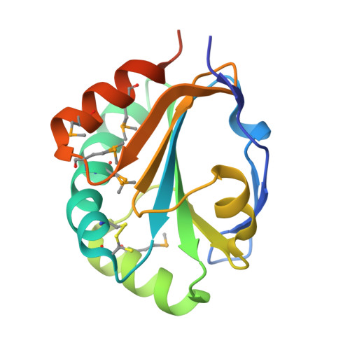
Mechanism of substrate specificity in Bacillus subtilis ResA, a thioredoxin-like protein involved in cytochrome c maturation
Colbert, C.L., Wu, Q., Erbel, P.J.A., Gardner, K.H., Deisenhofer, J.(2006) Proc Natl Acad Sci U S A 103: 4410-4415
- PubMed: 16537372
- DOI: https://doi.org/10.1073/pnas.0600552103
- Primary Citation of Related Structures:
2F9S - PubMed Abstract:
The covalent attachment of heme cofactors to the apo-polypeptides via thioether bonds is unique to the maturation of c-type cytochromes. A number of thiol-disulfide oxidoreductases prepare the apocytochrome for heme insertion in system I and II cytochrome c maturation. Although most thiol-disulfide oxidoreductases are nonspecific, the less common, specific thiol-disulfide oxidoreductases may be key to directing the usage of electrons. Here we demonstrate that unlike other thiol-disulfide oxidoreductases, the protein responsible for reducing oxidized apocytochrome c in Bacillus subtilis, ResA, is specific for cytochrome c550 and utilizes alternate conformations to recognize redox partners. We report solution NMR evidence that ResA undergoes a redox-dependent conformational change between oxidation states, as well as data showing that ResA utilizes a surface cavity present only in the reduced state to recognize a peptide derived from cytochrome c550. Finally, we confirm that ResA is a specific thiol-disulfide oxidoreductase by comparing its reactivity to our mimetic peptide with its reactivity to oxidized glutathione, a nonspecific substrate. This study biochemically demonstrates the specificity of this thiol-disulfide oxidoreductase and enables us to outline a structural mechanism of regulating the usage of electrons in a thiol-disulfide oxidoreductase system.
Organizational Affiliation:
Howard Hughes Medical Institute, and Department of Biochemistry, University of Texas Southwestern Medical Center, 6001 Forest Park Road, Dallas, TX 75390, USA.


