2F1K

Crystal structure of Synechocystis arogenate dehydrogenase


Experimental Data Snapshot

  • Method: X-RAY DIFFRACTION
  • Resolution: 1.55 Å
  • R-Value Free: 0.222 
  • R-Value Work: 0.189 
  • R-Value Observed: 0.190 

wwPDB Validation   3D Report Full Report


Ligand Structure Quality Assessment 


This is version 1.3 of the entry. See complete history


Literature

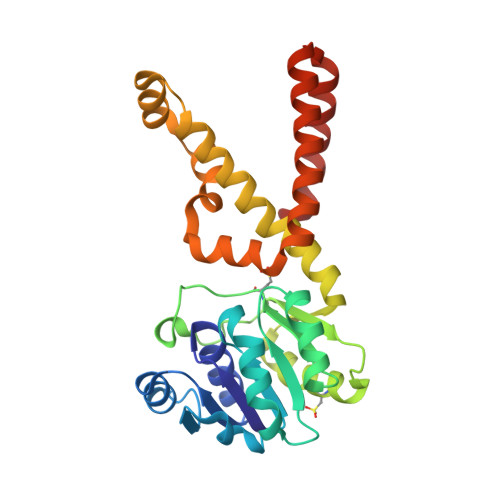
Biochemical Characterization and Crystal Structure of Synechocystis Arogenate Dehydrogenase Provide Insights into Catalytic Reaction

Legrand, P.Dumas, R.Seux, M.Rippert, P.Ravelli, R.Ferrer, J.-L.Matringe, M.

(2006) Structure 14: 767-776

  • DOI: https://doi.org/10.1016/j.str.2006.01.006
  • Primary Citation of Related Structures:  
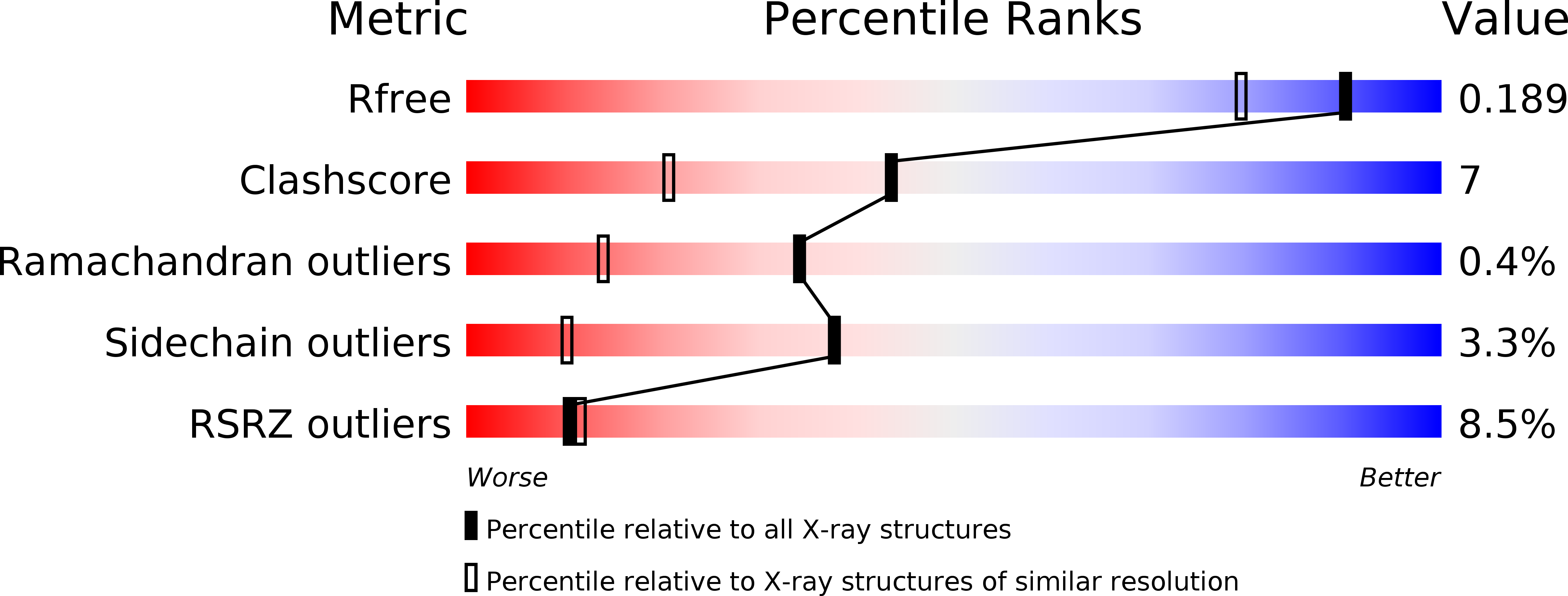
    2F1K

  • PubMed Abstract: 

    The extreme diversity in substrate specificity, and in the regulation mechanism of arogenate/prephenate dehydrogenase enzymes in nature, makes a comparative structural study of these enzymes of great interest. We report here on the biochemical and structural characterization of arogenate dehydrogenase from Synechocystis sp. (TyrAsy). This work paves the way for the understanding of the structural determinants leading to diversity in substrate specificity, and of the regulation mechanisms of arogenate/prephenate dehydrogenases. The overall structure of TyrAsy in complex with NADP was refined to 1.6 A. The asymmetric unit contains two TyrAsy homodimers, with each monomer consisting of a nucleotide binding N-terminal domain and a particularly unique alpha-helical C-terminal dimerization domain. The substrate arogenate was modeled into the active site. The model of the ternary complex enzyme-NADP-arogenate nicely reveals at the atomic level the concerted mechanism of the arogenate/prephenate dehydrogenase reaction.


  • Organizational Affiliation

    Institut de Biologie Structurale Jean-Pierre Ebel, Centre National de la Recherche Scientifique, Université Joseph Fourier, Laboratoire de Cristallographie et Cristallogenèse des Protéines/Groupe Synchrotron, 38027 Grenoble cedex 1, France.


Macromolecules
Find similar proteins by:  (by identity cutoff)  |  3D Structure
Entity ID: 1
MoleculeChains Sequence LengthOrganismDetailsImage
prephenate dehydrogenase
A, B, C, D
279Synechocystis sp. PCC 6803Mutation(s): 2 
Gene Names: D90910.1
EC: 1.3.1.43
UniProt
Find proteins for P73906 (Synechocystis sp. (strain PCC 6803 / Kazusa))
Explore P73906 
Go to UniProtKB:  P73906
Entity Groups  
Sequence Clusters30% Identity50% Identity70% Identity90% Identity95% Identity100% Identity
UniProt GroupP73906
Sequence Annotations
Expand
  • Reference Sequence
Experimental Data & Validation

Experimental Data

  • Method: X-RAY DIFFRACTION
  • Resolution: 1.55 Å
  • R-Value Free: 0.222 
  • R-Value Work: 0.189 
  • R-Value Observed: 0.190 
  • Space Group: P 1 21 1
Unit Cell:
Length ( Å )Angle ( ˚ )
a = 66.15α = 90
b = 70.85β = 90.3
c = 104.49γ = 90
Software Package:
Software NamePurpose
REFMACrefinement
ProDCdata collection
XDSdata scaling
SHARPphasing

Structure Validation

View Full Validation Report


Ligand Structure Quality Assessment 


Entry History 

Deposition Data

Revision History  (Full details and data files)

  • Version 1.0: 2006-05-09
    Type: Initial release
  • Version 1.1: 2008-05-01
    Changes: Version format compliance
  • Version 1.2: 2011-07-13
    Changes: Advisory, Source and taxonomy, Version format compliance
  • Version 1.3: 2017-10-18
    Changes: Advisory, Refinement description