2F1C

Crystal structure of the monomeric porin OmpG


Experimental Data Snapshot

  • Method: X-RAY DIFFRACTION
  • Resolution: 2.30 Å
  • R-Value Free: 0.260 
  • R-Value Work: 0.233 
  • R-Value Observed: 0.237 

wwPDB Validation   3D Report Full Report


Ligand Structure Quality Assessment 


This is version 1.3 of the entry. See complete history


Literature

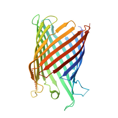
Crystal Structure of the Monomeric Porin OmpG.

Subbarao, G.V.van den Berg, B.

(2006) J Mol Biol 360: 750-759

  • DOI: https://doi.org/10.1016/j.jmb.2006.05.045
  • Primary Citation of Related Structures:  
    2F1C

  • PubMed Abstract: 

    The outer membrane (OM) of Gram-negative bacteria contains a large number of channel proteins that mediate the uptake of ions and nutrients necessary for growth and functioning of the cell. An important group of OM channel proteins are the porins, which mediate the non-specific, diffusion-based passage of small (<600 Da) polar molecules. All porins of Gram-negative bacteria that have been crystallized to date form stable trimers, with each monomer composed of a 16-stranded beta-barrel with a relatively narrow central pore. In contrast, the OmpG porin is unique, as it appears to function as a monomer. We have determined the X-ray crystal structure of OmpG from Escherichia coli to a resolution of 2.3 A. The structure shows a 14-stranded beta-barrel with a relatively simple architecture. Due to the absence of loops that fold back into the channel, OmpG has a large ( approximately 13 A) central pore that is considerably wider than those of other E. coli porins, and very similar in size to that of the toxin alpha-hemolysin. The architecture of the channel, together with previous biochemical and other data, suggests that OmpG may form a non-specific channel for the transport of larger oligosaccharides. The structure of OmpG provides the starting point for engineering studies aiming to generate selective channels and for the development of biosensors.


  • Organizational Affiliation

    Program in Molecular Medicine, University of Massachusetts Medical School, 373 Plantation Street, Worcester, MA 01605, USA.


Macromolecules
Find similar proteins by:  (by identity cutoff)  |  3D Structure
Entity ID: 1
MoleculeChains Sequence LengthOrganismDetailsImage
Outer membrane protein GA [auth X]286Escherichia coli K-12Mutation(s): 0 
Gene Names: ompG
Membrane Entity: Yes 
UniProt
Find proteins for P76045 (Escherichia coli (strain K12))
Explore P76045 
Go to UniProtKB:  P76045
Entity Groups  
Sequence Clusters30% Identity50% Identity70% Identity90% Identity95% Identity100% Identity
UniProt GroupP76045
Sequence Annotations
Expand
  • Reference Sequence
Small Molecules
Ligands 1 Unique
IDChains Name / Formula / InChI Key2D Diagram3D Interactions
C8E
Query on C8E

Download Ideal Coordinates CCD File 
B [auth X],
C [auth X]
(HYDROXYETHYLOXY)TRI(ETHYLOXY)OCTANE
C16 H34 O5
FEOZZFHAVXYAMB-UHFFFAOYSA-N
Experimental Data & Validation

Experimental Data

  • Method: X-RAY DIFFRACTION
  • Resolution: 2.30 Å
  • R-Value Free: 0.260 
  • R-Value Work: 0.233 
  • R-Value Observed: 0.237 
  • Space Group: C 1 2 1
Unit Cell:
Length ( Å )Angle ( ˚ )
a = 98.64α = 90
b = 70.142β = 104.06
c = 59.501γ = 90
Software Package:
Software NamePurpose
HKL-2000data collection
SCALEPACKdata scaling
SHARPphasing
CNSrefinement
HKL-2000data reduction

Structure Validation

View Full Validation Report


Ligand Structure Quality Assessment 


Entry History 

Deposition Data

Revision History  (Full details and data files)

  • Version 1.0: 2006-06-27
    Type: Initial release
  • Version 1.1: 2008-05-01
    Changes: Version format compliance
  • Version 1.2: 2011-07-13
    Changes: Derived calculations, Version format compliance
  • Version 1.3: 2024-02-14
    Changes: Data collection, Database references, Derived calculations