2EX3

Bacteriophage phi29 DNA polymerase bound to terminal protein


Experimental Data Snapshot

  • Method: X-RAY DIFFRACTION
  • Resolution: 3.00 Å
  • R-Value Free: 0.229 
  • R-Value Work: 0.200 
  • R-Value Observed: 0.203 

wwPDB Validation   3D Report Full Report


This is version 1.6 of the entry. See complete history


Literature

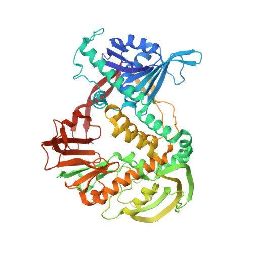
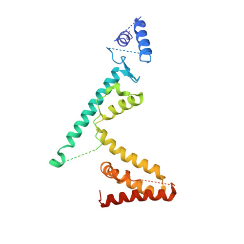
The phi29 DNA polymerase:protein-primer structure suggests a model for the initiation to elongation transition

Kamtekar, S.Berman, A.J.Wang, J.Lazaro, J.M.de Vega, M.Blanco, L.Salas, M.Steitz, T.A.

(2006) EMBO J 25: 1335-1343

  • DOI: https://doi.org/10.1038/sj.emboj.7601027
  • Primary Citation of Related Structures:  
    2EX3

  • PubMed Abstract: 

    The absolute requirement for primers in the initiation of DNA synthesis poses a problem for replicating the ends of linear chromosomes. The DNA polymerase of bacteriophage phi29 solves this problem by using a serine hydroxyl of terminal protein to prime replication. The 3.0 A resolution structure shows one domain of terminal protein making no interactions, a second binding the polymerase and a third domain containing the priming serine occupying the same binding cleft in the polymerase as duplex DNA does during elongation. Thus, the progressively elongating DNA duplex product must displace this priming domain. Further, this heterodimer of polymerase and terminal protein cannot accommodate upstream template DNA, thereby explaining its specificity for initiating DNA synthesis only at the ends of the bacteriophage genome. We propose a model for the transition from the initiation to the elongation phases in which the priming domain of terminal protein moves out of the active site as polymerase elongates the primer strand. The model indicates that terminal protein should dissociate from polymerase after the incorporation of approximately six nucleotides.


  • Organizational Affiliation

    Department of Molecular Biophysics and Biochemistry, Yale University, New Haven, CT 06520-8114, USA.


Macromolecules
Find similar proteins by:  (by identity cutoff)  |  3D Structure
Entity ID: 1
MoleculeChains Sequence LengthOrganismDetailsImage
DNA polymerase
A, C, E, G, I
A, C, E, G, I, K
575Salasvirus phi29Mutation(s): 2 
Gene Names: 2
EC: 2.7.7.7
UniProt
Find proteins for P03680 (Bacillus phage phi29)
Explore P03680 
Go to UniProtKB:  P03680
Entity Groups  
Sequence Clusters30% Identity50% Identity70% Identity90% Identity95% Identity100% Identity
UniProt GroupP03680
Sequence Annotations
Expand
  • Reference Sequence
Find similar proteins by:  (by identity cutoff)  |  3D Structure
Entity ID: 2
MoleculeChains Sequence LengthOrganismDetailsImage
DNA terminal protein
B, D, F, H, J
B, D, F, H, J, L
230Salasvirus phi29Mutation(s): 0 
Gene Names: 3
UniProt
Find proteins for P03681 (Bacillus phage phi29)
Explore P03681 
Go to UniProtKB:  P03681
Entity Groups  
Sequence Clusters30% Identity50% Identity70% Identity90% Identity95% Identity100% Identity
UniProt GroupP03681
Sequence Annotations
Expand
  • Reference Sequence
Small Molecules
Ligands 1 Unique
IDChains Name / Formula / InChI Key2D Diagram3D Interactions
PB
Query on PB

Download Ideal Coordinates CCD File 
AA [auth K]
BA [auth K]
M [auth A]
N [auth A]
O [auth A]
AA [auth K],
BA [auth K],
M [auth A],
N [auth A],
O [auth A],
P [auth C],
Q [auth C],
R [auth C],
S [auth E],
T [auth E],
U [auth E],
V [auth G],
W [auth G],
X [auth G],
Y [auth I],
Z [auth I]
LEAD (II) ION
Pb
RVPVRDXYQKGNMQ-UHFFFAOYSA-N
Experimental Data & Validation

Experimental Data

  • Method: X-RAY DIFFRACTION
  • Resolution: 3.00 Å
  • R-Value Free: 0.229 
  • R-Value Work: 0.200 
  • R-Value Observed: 0.203 
  • Space Group: C 1 2 1
Unit Cell:
Length ( Å )Angle ( ˚ )
a = 304.933α = 90
b = 220.281β = 45.4
c = 217.165γ = 90
Software Package:
Software NamePurpose
DENZOdata reduction
SCALEPACKdata scaling
REFMACrefinement
PDB_EXTRACTdata extraction
HKL-2000data reduction
PHASERphasing

Structure Validation

View Full Validation Report


Entry History 

Deposition Data

Revision History  (Full details and data files)

  • Version 1.0: 2006-03-14
    Type: Initial release
  • Version 1.1: 2008-05-01
    Changes: Version format compliance
  • Version 1.2: 2011-07-13
    Changes: Advisory, Version format compliance
  • Version 1.3: 2017-10-18
    Changes: Refinement description
  • Version 1.4: 2021-10-20
    Changes: Database references, Derived calculations
  • Version 1.5: 2024-02-14
    Changes: Data collection, Refinement description
  • Version 1.6: 2024-04-03
    Changes: Refinement description