2ESB

Crystal structure of human DUSP18


Experimental Data Snapshot

  • Method: X-RAY DIFFRACTION
  • Resolution: 2.00 Å
  • R-Value Free: 0.189 
  • R-Value Work: 0.159 
  • R-Value Observed: 0.161 

wwPDB Validation   3D Report Full Report


This is version 1.3 of the entry. See complete history


Literature

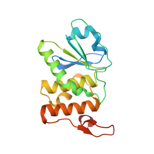
Structure of human DSP18, a member of the dual-specificity protein tyrosine phosphatase family.

Jeong, D.G.Cho, Y.H.Yoon, T.S.Kim, J.H.Son, J.H.Ryu, S.E.Kim, S.J.

(2006) Acta Crystallogr D Biol Crystallogr 62: 582-588

  • DOI: https://doi.org/10.1107/S0907444906010109
  • Primary Citation of Related Structures:  
    2ESB

  • PubMed Abstract: 

    The human dual-specificity protein phosphatase 18 (DSP18) gene and its protein product have recently been characterized. Like most DSPs, DSP18 displays dephosphorylating activity towards both phosphotyrosine and phosphothreonine residues. However, DSP18 is distinct from other known DSPs in terms of the existence of approximately 30 residues at the C-terminus of the catalytic domain and an unusual optimum activity profile at 328 K. The crystal structure of human DSP18 has been determined at 2.0 A resolution. The catalytic domain of DSP18 adopts a fold similar to that known for other DSP structures. Although good alignments are found with other DSPs, substantial differences are also found in the regions surrounding the active site, suggesting that DSP18 constitutes a unique structure with a distinct substrate specificity. Furthermore, the residues at the C-terminus fold into two antiparallel beta-strands and participate in extensive interactions with the catalytic domain, explaining the thermostability of DSP18.


  • Organizational Affiliation

    Systemic Proteomics Research Center, Korea Research Institute of Bioscience and Biotechnology, 52 Eoeun-Dong, Yuseong-Gu, Daejeon 305-333, South Korea.


Macromolecules
Find similar proteins by:  (by identity cutoff)  |  3D Structure
Entity ID: 1
MoleculeChains Sequence LengthOrganismDetailsImage
Dual specificity protein phosphatase 18188Homo sapiensMutation(s): 0 
EC: 3.1.3.48 (PDB Primary Data), 3.1.3.16 (PDB Primary Data)
UniProt & NIH Common Fund Data Resources
Find proteins for Q8NEJ0 (Homo sapiens)
Explore Q8NEJ0 
Go to UniProtKB:  Q8NEJ0
PHAROS:  Q8NEJ0
GTEx:  ENSG00000167065 
Entity Groups  
Sequence Clusters30% Identity50% Identity70% Identity90% Identity95% Identity100% Identity
UniProt GroupQ8NEJ0
Sequence Annotations
Expand
  • Reference Sequence
Small Molecules
Ligands 2 Unique
IDChains Name / Formula / InChI Key2D Diagram3D Interactions
EPE
Query on EPE

Download Ideal Coordinates CCD File 
C [auth A]4-(2-HYDROXYETHYL)-1-PIPERAZINE ETHANESULFONIC ACID
C8 H18 N2 O4 S
JKMHFZQWWAIEOD-UHFFFAOYSA-N
ACT
Query on ACT

Download Ideal Coordinates CCD File 
B [auth A]ACETATE ION
C2 H3 O2
QTBSBXVTEAMEQO-UHFFFAOYSA-M
Experimental Data & Validation

Experimental Data

  • Method: X-RAY DIFFRACTION
  • Resolution: 2.00 Å
  • R-Value Free: 0.189 
  • R-Value Work: 0.159 
  • R-Value Observed: 0.161 
  • Space Group: C 2 2 21
Unit Cell:
Length ( Å )Angle ( ˚ )
a = 35.74α = 90
b = 96.083β = 90
c = 116.654γ = 90
Software Package:
Software NamePurpose
HKL-2000data collection
SCALEPACKdata scaling
EPMRphasing
CNSrefinement
HKL-2000data reduction

Structure Validation

View Full Validation Report


Entry History 

Deposition Data

Revision History  (Full details and data files)

  • Version 1.0: 2006-06-06
    Type: Initial release
  • Version 1.1: 2008-05-01
    Changes: Version format compliance
  • Version 1.2: 2011-07-13
    Changes: Version format compliance
  • Version 1.3: 2024-03-13
    Changes: Data collection, Database references, Derived calculations