


Crystal Structure Of Uroporphyrinogen Decarboxylase From Aquifex aeolicus
Bagautdinov, B., Kunishima, N.To be published.
Experimental Data Snapshot
wwPDB Validation 3D Report Full Report

Entity ID: 1 | |||||
|---|---|---|---|---|---|
| Molecule | Chains | Sequence Length | Organism | Details | Image |
| Uroporphyrinogen decarboxylase | 338 | Aquifex aeolicus | Mutation(s): 0 EC: 4.1.1.37 | 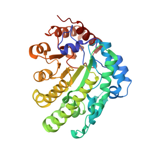 | |
UniProt | |||||
Find proteins for O66667 (Aquifex aeolicus (strain VF5)) Explore O66667 Go to UniProtKB: O66667 | |||||
Entity Groups | |||||
| Sequence Clusters | 30% Identity50% Identity70% Identity90% Identity95% Identity100% Identity | ||||
| UniProt Group | O66667 | ||||
Sequence AnnotationsExpand | |||||
| |||||
| Ligands 1 Unique | |||||
|---|---|---|---|---|---|
| ID | Chains | Name / Formula / InChI Key | 2D Diagram | 3D Interactions | |
| ACT Query on ACT | C [auth A] | ACETATE ION C2 H3 O2 QTBSBXVTEAMEQO-UHFFFAOYSA-M |  | ||
| Length ( Å ) | Angle ( ˚ ) |
|---|---|
| a = 65.473 | α = 90 |
| b = 80.403 | β = 90 |
| c = 128.999 | γ = 90 |
| Software Name | Purpose |
|---|---|
| HKL-2000 | data collection |
| AMoRE | phasing |
| CNS | refinement |
| HKL-2000 | data reduction |
| SCALEPACK | data scaling |
RCSB PDB (citation) is hosted by
RCSB PDB is a member of the

