


Crystal Structure of a Putative protein (AQ1627) from Aquifex aeolicus
Kumarevel, T.S., Karthe, P., Nakano, N., Kuramitsu, S., Yokoyama, S.To be published.
Experimental Data Snapshot
wwPDB Validation 3D Report Full Report

Entity ID: 1 | |||||
|---|---|---|---|---|---|
| Molecule | Chains | Sequence Length | Organism | Details | Image |
| aq_1627 protein | 126 | Aquifex aeolicus | Mutation(s): 0 | 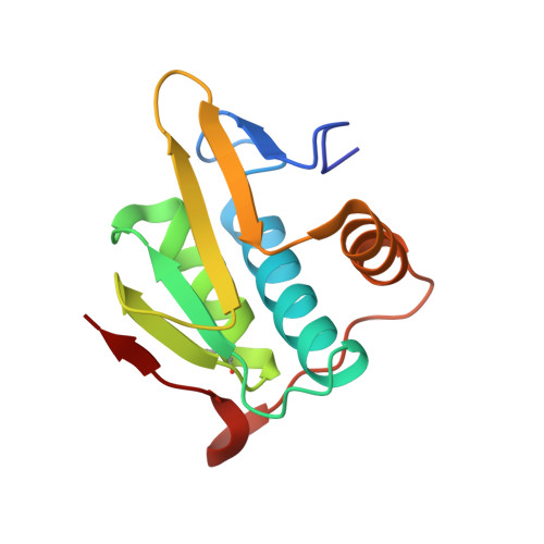 | |
UniProt | |||||
Find proteins for O67549 (Aquifex aeolicus (strain VF5)) Explore O67549 Go to UniProtKB: O67549 | |||||
Entity Groups | |||||
| Sequence Clusters | 30% Identity50% Identity70% Identity90% Identity95% Identity100% Identity | ||||
| UniProt Group | O67549 | ||||
Sequence AnnotationsExpand | |||||
| |||||
| Modified Residues 1 Unique | |||||
|---|---|---|---|---|---|
| ID | Chains | Type | Formula | 2D Diagram | Parent |
| MSE Query on MSE | A, B | L-PEPTIDE LINKING | C5 H11 N O2 Se |  | MET |
| Length ( Å ) | Angle ( ˚ ) |
|---|---|
| a = 73.353 | α = 90 |
| b = 53.873 | β = 96.09 |
| c = 60.168 | γ = 90 |
| Software Name | Purpose |
|---|---|
| CNS | refinement |
| BSS | data collection |
| HKL-2000 | data reduction |
| HKL-2000 | data scaling |
| MOLREP | phasing |
RCSB PDB (citation) is hosted by
RCSB PDB is a member of the

