


Crystal structure of the 3-dehydroquinate dehydratase from Aquifex aeolicus VF5
Karthe, P., Kumarevel, T.S., Ebihara, A., Kuramitsu, S., Yokoyama, S.To be published.
Experimental Data Snapshot
wwPDB Validation 3D Report Full Report

Entity ID: 1 | |||||
|---|---|---|---|---|---|
| Molecule | Chains | Sequence Length | Organism | Details | Image |
| 3-dehydroquinate dehydratase | A, B [auth C] | 219 | Aquifex aeolicus | Mutation(s): 0 Gene Names: aroD EC: 4.2.1.10 | 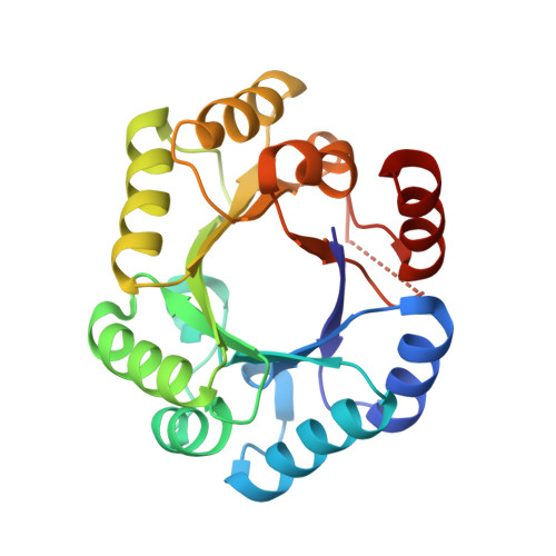 |
UniProt | |||||
Find proteins for O66440 (Aquifex aeolicus (strain VF5)) Explore O66440 Go to UniProtKB: O66440 | |||||
Entity Groups | |||||
| Sequence Clusters | 30% Identity50% Identity70% Identity90% Identity95% Identity100% Identity | ||||
| UniProt Group | O66440 | ||||
Sequence AnnotationsExpand | |||||
| |||||
| Ligands 1 Unique | |||||
|---|---|---|---|---|---|
| ID | Chains | Name / Formula / InChI Key | 2D Diagram | 3D Interactions | |
| TLA Query on TLA | C [auth A], D [auth C] | L(+)-TARTARIC ACID C4 H6 O6 FEWJPZIEWOKRBE-JCYAYHJZSA-N |  | ||
| Length ( Å ) | Angle ( ˚ ) |
|---|---|
| a = 52.937 | α = 90 |
| b = 67.614 | β = 99.2 |
| c = 60.238 | γ = 90 |
| Software Name | Purpose |
|---|---|
| CNS | refinement |
| BSS | data collection |
| HKL-2000 | data reduction |
| HKL-2000 | data scaling |
| SOLVE | phasing |
RCSB PDB (citation) is hosted by
RCSB PDB is a member of the

