2EC6

Placopecten Striated Muscle Myosin II


Experimental Data Snapshot

  • Method: X-RAY DIFFRACTION
  • Resolution: 3.25 Å
  • R-Value Free: 0.300 
  • R-Value Work: 0.279 
  • R-Value Observed: 0.279 

wwPDB Validation   3D Report Full Report


This is version 1.3 of the entry. See complete history


Literature

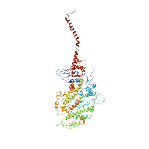
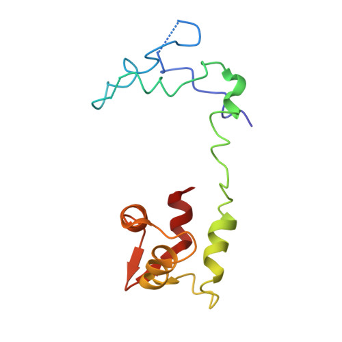
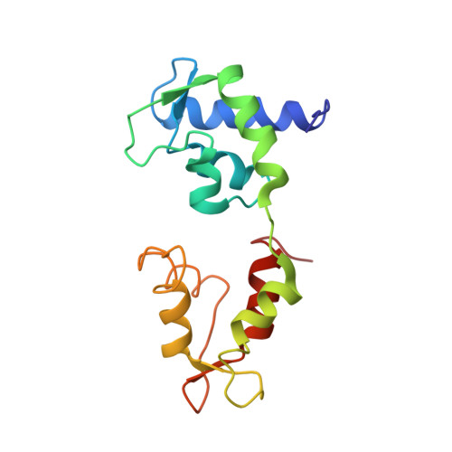
Rigor-like structures from muscle myosins reveal key mechanical elements in the transduction pathways of this allosteric motor.

Yang, Y.Gourinath, S.Kovacs, M.Nyitray, L.Reutzel, R.Himmel, D.M.O'Neall-Hennessey, E.Reshetnikova, L.Szent-Gyorgyi, A.G.Brown, J.H.Cohen, C.

(2007) Structure 15: 553-564

  • DOI: https://doi.org/10.1016/j.str.2007.03.010
  • Primary Citation of Related Structures:  
    2EC6, 2OS8, 2OTG, 3I5F, 3I5G, 3I5H, 3I5I

  • PubMed Abstract: 

    Unlike processive cellular motors such as myosin V, whose structure has recently been determined in a "rigor-like" conformation, myosin II from contracting muscle filaments necessarily spends most of its time detached from actin. By using squid and sea scallop sources, however, we have now obtained similar rigor-like atomic structures for muscle myosin heads (S1). The significance of the hallmark closed actin-binding cleft in these crystal structures is supported here by actin/S1-binding studies. These structures reveal how different duty ratios, and hence cellular functions, of the myosin isoforms may be accounted for, in part, on the basis of detailed differences in interdomain contacts. Moreover, the rigor-like position of switch II turns out to be unique for myosin V. The overall arrangements of subdomains in the motor are relatively conserved in each of the known contractile states, and we explore qualitatively the energetics of these states.


  • Organizational Affiliation

    Rosenstiel Basic Medical Sciences Research Center, Brandeis University, Waltham, MA 02454, USA.


Macromolecules
Find similar proteins by:  (by identity cutoff)  |  3D Structure
Entity ID: 1
MoleculeChains Sequence LengthOrganismDetailsImage
Myosin heavy chain838Placopecten magellanicusMutation(s): 0 
UniProt
Find proteins for Q26079 (Placopecten magellanicus)
Explore Q26079 
Go to UniProtKB:  Q26079
Entity Groups  
Sequence Clusters30% Identity50% Identity70% Identity90% Identity95% Identity100% Identity
UniProt GroupQ26079
Sequence Annotations
Expand
  • Reference Sequence
Find similar proteins by:  (by identity cutoff)  |  3D Structure
Entity ID: 2
MoleculeChains Sequence LengthOrganismDetailsImage
Myosin regulatory light chain133Placopecten magellanicusMutation(s): 0 
UniProt
Find proteins for Q26069 (Placopecten magellanicus)
Explore Q26069 
Go to UniProtKB:  Q26069
Entity Groups  
Sequence Clusters30% Identity50% Identity70% Identity90% Identity95% Identity100% Identity
UniProt GroupQ26069
Sequence Annotations
Expand
  • Reference Sequence
Find similar proteins by:  (by identity cutoff)  |  3D Structure
Entity ID: 3
MoleculeChains Sequence LengthOrganismDetailsImage
Myosin essential light chain156Placopecten magellanicusMutation(s): 2 
UniProt
Find proteins for Q26066 (Placopecten magellanicus)
Explore Q26066 
Go to UniProtKB:  Q26066
Entity Groups  
Sequence Clusters30% Identity50% Identity70% Identity90% Identity95% Identity100% Identity
UniProt GroupQ26066
Sequence Annotations
Expand
  • Reference Sequence
Small Molecules
Ligands 1 Unique
IDChains Name / Formula / InChI Key2D Diagram3D Interactions
CA
Query on CA

Download Ideal Coordinates CCD File 
D [auth C]CALCIUM ION
Ca
BHPQYMZQTOCNFJ-UHFFFAOYSA-N
Experimental Data & Validation

Experimental Data

  • Method: X-RAY DIFFRACTION
  • Resolution: 3.25 Å
  • R-Value Free: 0.300 
  • R-Value Work: 0.279 
  • R-Value Observed: 0.279 
  • Space Group: P 1 21 1
Unit Cell:
Length ( Å )Angle ( ˚ )
a = 85.273α = 90
b = 50.366β = 101.04
c = 156.774γ = 90
Software Package:
Software NamePurpose
ADSCdata collection
AMoREphasing
CNSrefinement
DENZOdata reduction
SCALEPACKdata scaling

Structure Validation

View Full Validation Report


Entry History 

Deposition Data

Revision History  (Full details and data files)

  • Version 1.0: 2008-02-26
    Type: Initial release
  • Version 1.1: 2011-07-13
    Changes: Derived calculations, Version format compliance
  • Version 1.2: 2018-08-08
    Changes: Data collection, Source and taxonomy, Structure summary
  • Version 1.3: 2021-11-10
    Changes: Database references, Derived calculations