2EB4

Crystal structure of apo-HpcG


Experimental Data Snapshot

  • Method: X-RAY DIFFRACTION
  • Resolution: 1.60 Å
  • R-Value Free: 0.229 
  • R-Value Work: 0.206 
  • R-Value Observed: 0.207 

wwPDB Validation   3D Report Full Report


This is version 1.3 of the entry. See complete history


Literature

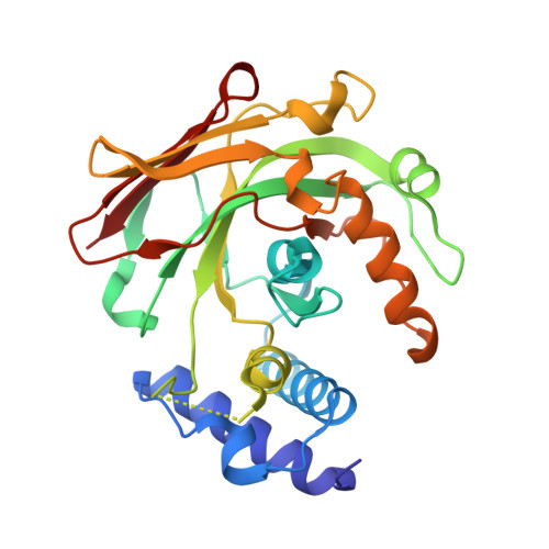
Structure and Mechanism of HpcG, a Hydratase in the Homoprotocatechuate Degradation Pathway of Escherichia coli

Izumi, A.Rea, D.Adachi, T.Unzai, S.Park, S.Y.Roper, D.I.Tame, J.R.H.

(2007) J Mol Biol 370: 899-911

  • DOI: https://doi.org/10.1016/j.jmb.2007.05.006
  • Primary Citation of Related Structures:  
    2EB4, 2EB5, 2EB6

  • PubMed Abstract: 

    HpcG catalyses the hydration of a carbon-carbon double bond without the aid of any cofactor other than a simple divalent metal ion such as Mg(2+). Since the substrate has a nearby carbonyl group, it is believed that it first isomerises to form a pair of conjugated double bonds in the enol tautomer before Michael addition of water. Previous chemical studies of the reaction, and that of the related enzyme MhpD, have failed to provide a clear picture of the mechanism. The substrate itself is unstable, preventing co-crystallisation or soaking of crystals, but oxalate is a strong competitive inhibitor. We have solved the crystal structure of the protein in the apo form, and with magnesium and oxalate bound. Modelling substrate into the active site suggests the attacking water molecule is not part of the metal coordination shell, in contrast to a previous proposal. Our model suggests that geometrically strained cis isomer intermediates do not lie on the reaction pathway, and that separate groups are involved in the isomerisation and hydration steps.


  • Organizational Affiliation

    Protein Design Laboratory, Yokohama City University, Suehiro 1-7-29, Tsurumi, Yokohama 230-0045, Japan.


Macromolecules
Find similar proteins by:  (by identity cutoff)  |  3D Structure
Entity ID: 1
MoleculeChains Sequence LengthOrganismDetailsImage
2-oxo-hept-3-ene-1,7-dioate hydratase
A, B, C, D, E
267Escherichia coliMutation(s): 0 
Gene Names: hpcG
EC: 4.2.1
UniProt
Find proteins for Q46982 (Escherichia coli)
Explore Q46982 
Go to UniProtKB:  Q46982
Entity Groups  
Sequence Clusters30% Identity50% Identity70% Identity90% Identity95% Identity100% Identity
UniProt GroupQ46982
Sequence Annotations
Expand
  • Reference Sequence
Small Molecules
Ligands 2 Unique
IDChains Name / Formula / InChI Key2D Diagram3D Interactions
SCN
Query on SCN

Download Ideal Coordinates CCD File 
F [auth A]
G [auth A]
H [auth A]
J [auth B]
K [auth B]
F [auth A],
G [auth A],
H [auth A],
J [auth B],
K [auth B],
M [auth C],
N [auth C],
P [auth D],
R [auth E]
THIOCYANATE ION
C N S
ZMZDMBWJUHKJPS-UHFFFAOYSA-M
NA
Query on NA

Download Ideal Coordinates CCD File 
I [auth A],
L [auth B],
O [auth C],
Q [auth D],
S [auth E]
SODIUM ION
Na
FKNQFGJONOIPTF-UHFFFAOYSA-N
Experimental Data & Validation

Experimental Data

  • Method: X-RAY DIFFRACTION
  • Resolution: 1.60 Å
  • R-Value Free: 0.229 
  • R-Value Work: 0.206 
  • R-Value Observed: 0.207 
  • Space Group: P 43 21 2
Unit Cell:
Length ( Å )Angle ( ˚ )
a = 136.74α = 90
b = 136.74β = 90
c = 192.882γ = 90
Software Package:
Software NamePurpose
REFMACrefinement
HKL-2000data collection
DENZOdata reduction
SCALEPACKdata scaling
SOLVEphasing

Structure Validation

View Full Validation Report


Entry History 

Deposition Data

Revision History  (Full details and data files)

  • Version 1.0: 2007-07-17
    Type: Initial release
  • Version 1.1: 2008-04-30
    Changes: Version format compliance
  • Version 1.2: 2011-07-13
    Changes: Version format compliance
  • Version 1.3: 2024-03-13
    Changes: Data collection, Database references, Derived calculations, Refinement description