


Crystal Structure Of Biotin Protein Ligase From Aquifex Aeolicus
Bagautdinov, B., Kunishima, N.To be published.
Experimental Data Snapshot
wwPDB Validation 3D Report Full Report

Entity ID: 1 | |||||
|---|---|---|---|---|---|
| Molecule | Chains | Sequence Length | Organism | Details | Image |
| Biotin [acetyl-CoA-carboxylase] ligase | 233 | Aquifex aeolicus | Mutation(s): 2 Gene Names: birA EC: 6.3.4.15 | 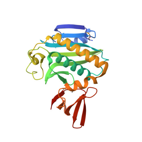 | |
UniProt | |||||
Find proteins for O66837 (Aquifex aeolicus (strain VF5)) Explore O66837 Go to UniProtKB: O66837 | |||||
Entity Groups | |||||
| Sequence Clusters | 30% Identity50% Identity70% Identity90% Identity95% Identity100% Identity | ||||
| UniProt Group | O66837 | ||||
Sequence AnnotationsExpand | |||||
| |||||
| Modified Residues 1 Unique | |||||
|---|---|---|---|---|---|
| ID | Chains | Type | Formula | 2D Diagram | Parent |
| MSE Query on MSE | A, B | L-PEPTIDE LINKING | C5 H11 N O2 Se |  | MET |
| Length ( Å ) | Angle ( ˚ ) |
|---|---|
| a = 55.076 | α = 90 |
| b = 60.589 | β = 91.99 |
| c = 73.106 | γ = 90 |
| Software Name | Purpose |
|---|---|
| HKL-2000 | data collection |
| AMoRE | phasing |
| CNS | refinement |
| HKL-2000 | data reduction |
| SCALEPACK | data scaling |
RCSB PDB (citation) is hosted by
RCSB PDB is a member of the

