2E5Y

Epsilon subunit and ATP complex of F1F0-ATP synthase from the Thermophilic Bacillus PS3


Experimental Data Snapshot

  • Method: X-RAY DIFFRACTION
  • Resolution: 1.92 Å
  • R-Value Free: 0.250 
  • R-Value Work: 0.200 
  • R-Value Observed: 0.202 

wwPDB Validation   3D Report Full Report


Ligand Structure Quality Assessment 


This is version 1.3 of the entry. See complete history


Literature

Structures of the thermophilic F1-ATPase {varepsilon} subunit suggesting ATP-regulated arm motion of its C-terminal domain in F1

Yagi, H.Kajiwara, N.Tanaka, H.Tsukihara, T.Kato-Yamada, Y.Yoshida, M.Akutsu, H.

(2007) Proc Natl Acad Sci U S A 104: 11233-11238

  • DOI: https://doi.org/10.1073/pnas.0701045104
  • Primary Citation of Related Structures:  
    2E5T, 2E5U, 2E5Y

  • PubMed Abstract: 

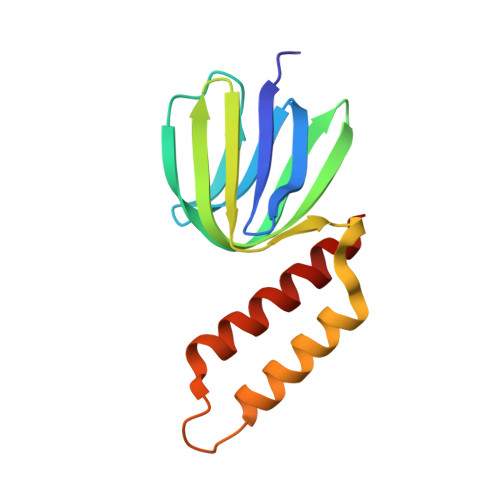
    The epsilon subunit of bacterial and chloroplast F(o)F(1)-ATP synthases modulates their ATP hydrolysis activity. Here, we report the crystal structure of the ATP-bound epsilon subunit from a thermophilic Bacillus PS3 at 1.9-A resolution. The C-terminal two alpha-helices were folded into a hairpin, sitting on the beta sandwich structure, as reported for Escherichia coli. A previously undescribed ATP binding motif, I(L)DXXRA, recognizes ATP together with three arginine and one glutamate residues. The E. coli epsilon subunit binds ATP in a similar manner, as judged on NMR. We also determined solution structures of the C-terminal domain of the PS3 epsilon subunit and relaxation parameters of the whole molecule by NMR. The two helices fold into a hairpin in the presence of ATP but extend in the absence of ATP. The latter structure has more helical regions and is much more flexible than the former. These results suggest that the epsilon C-terminal domain can undergo an arm-like motion in response to an ATP concentration change and thereby contribute to regulation of F(o)F(1)-ATP synthase.


  • Organizational Affiliation

    Institute for Protein Research, Osaka University, 3-2 Yamadaoka, Suita 565-0871, Japan.


Macromolecules
Find similar proteins by:  (by identity cutoff)  |  3D Structure
Entity ID: 1
MoleculeChains Sequence LengthOrganismDetailsImage
ATP synthase epsilon chain
A, B
133Bacillus sp. PS3Mutation(s): 0 
EC: 3.6.3.14
UniProt
Find proteins for P07678 (Bacillus sp. (strain PS3))
Explore P07678 
Go to UniProtKB:  P07678
Entity Groups  
Sequence Clusters30% Identity50% Identity70% Identity90% Identity95% Identity100% Identity
UniProt GroupP07678
Sequence Annotations
Expand
  • Reference Sequence
Small Molecules
Ligands 1 Unique
IDChains Name / Formula / InChI Key2D Diagram3D Interactions
ATP
Query on ATP

Download Ideal Coordinates CCD File 
C [auth A],
D [auth B]
ADENOSINE-5'-TRIPHOSPHATE
C10 H16 N5 O13 P3
ZKHQWZAMYRWXGA-KQYNXXCUSA-N
Binding Affinity Annotations 
IDSourceBinding Affinity
ATP Binding MOAD:  2E5Y Kd: 2.20e+7 (nM) from 1 assay(s)
PDBBind:  2E5Y Kd: 1400 (nM) from 1 assay(s)
Experimental Data & Validation

Experimental Data

  • Method: X-RAY DIFFRACTION
  • Resolution: 1.92 Å
  • R-Value Free: 0.250 
  • R-Value Work: 0.200 
  • R-Value Observed: 0.202 
  • Space Group: P 21 21 21
Unit Cell:
Length ( Å )Angle ( ˚ )
a = 37.48α = 90
b = 64.917β = 90
c = 103.944γ = 90
Software Package:
Software NamePurpose
REFMACrefinement
MOSFLMdata reduction
SCALAdata scaling

Structure Validation

View Full Validation Report


Ligand Structure Quality Assessment 


Entry History 

Deposition Data

Revision History  (Full details and data files)

  • Version 1.0: 2007-07-10
    Type: Initial release
  • Version 1.1: 2008-04-30
    Changes: Version format compliance
  • Version 1.2: 2011-07-13
    Changes: Version format compliance
  • Version 1.3: 2023-10-25
    Changes: Data collection, Database references, Derived calculations, Refinement description