2E1R

Structure of eEF2 in complex with a sordarin derivative


Experimental Data Snapshot

  • Method: X-RAY DIFFRACTION
  • Resolution: 3.15 Å
  • R-Value Free: 0.270 
  • R-Value Work: 0.220 
  • R-Value Observed: 0.224 

wwPDB Validation   3D Report Full Report


Ligand Structure Quality Assessment 


This is version 1.4 of the entry. See complete history


Literature

Sordarin derivatives induce a novel conformation of the yeast ribosome translocation factor eEF2

Soe, R.Mosley, R.T.Justice, M.Nielsen-Kahn, J.Shastry, M.Merrill, A.R.Andersen, G.R.

(2007) J Biol Chem 282: 657-666

  • DOI: https://doi.org/10.1074/jbc.M607830200
  • Primary Citation of Related Structures:  
    2E1R, 2NPF

  • PubMed Abstract: 

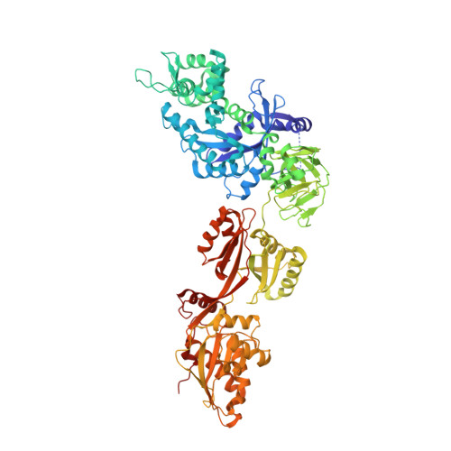
    The sordarins are fungal specific inhibitors of the translation factor eEF2, which catalyzes the translocation of tRNA and mRNA after peptide bond formation. We have determined the crystal structures of eEF2 in complex with two novel sordarin derivatives. In both structures, the three domains of eEF2 that form the ligand-binding pocket are oriented in a different manner relative to the rest of eEF2 compared with our previous structure of eEF2 in complex with the parent natural product sordarin. Yeast eEF2 is also shown to bind adenylic nucleotides, which can be displaced by sordarin, suggesting that ADP or ATP also bind to the three C-terminal domains of eEF2. Fusidic acid is a universal inhibitor of translation that targets EF-G or eEF2 and is widely used as an antibiotic against Gram-positive bacteria. Based on mutations conferring resistance to fusidic acid, cryo-EM reconstructions, and x-ray structures of eEF2, EF-G, and an EF-G homolog, we suggest that the conformation of EF-G stalled on the 70 S ribosome by fusidic acid is similar to that of eEF2 trapped on the 80 S ribosome by sordarin.


  • Organizational Affiliation

    Centre for Structural Biology, Department of Molecular Biology, University of Aarhus, DK-8000 Aarhus C, Denmark.


Macromolecules
Find similar proteins by:  (by identity cutoff)  |  3D Structure
Entity ID: 1
MoleculeChains Sequence LengthOrganismDetailsImage
Elongation factor 2842Saccharomyces cerevisiaeMutation(s): 0 
UniProt
Find proteins for P32324 (Saccharomyces cerevisiae (strain ATCC 204508 / S288c))
Explore P32324 
Go to UniProtKB:  P32324
Entity Groups  
Sequence Clusters30% Identity50% Identity70% Identity90% Identity95% Identity100% Identity
UniProt GroupP32324
Sequence Annotations
Expand
  • Reference Sequence
Small Molecules
Ligands 2 Unique
IDChains Name / Formula / InChI Key2D Diagram3D Interactions
SOD
Query on SOD

Download Ideal Coordinates CCD File 
B [auth A](1S,4S,5S,6R,9S,11S)-6-CHLORO-9-FORMYL-13-ISOPROPYL-5-METHYL-2-({[(3AR,5R,7R ,7AS)-7-METHYL-3-METHYLENEHEXAHYDRO-2H-FURO[2,3-C]PYRAN-5-YL]OXY}METHYL)TETR ACYCLO[7.4.0.02,11.04,8]TRIDEC-12-ENE-1-CARBOXYLIC ACI
C29 H39 Cl O6
OFLZRRPTCKISFB-SHWNUAPTSA-N
GDP
Query on GDP

Download Ideal Coordinates CCD File 
C [auth A]GUANOSINE-5'-DIPHOSPHATE
C10 H15 N5 O11 P2
QGWNDRXFNXRZMB-UUOKFMHZSA-N
Experimental Data & Validation

Experimental Data

  • Method: X-RAY DIFFRACTION
  • Resolution: 3.15 Å
  • R-Value Free: 0.270 
  • R-Value Work: 0.220 
  • R-Value Observed: 0.224 
  • Space Group: P 21 21 2
Unit Cell:
Length ( Å )Angle ( ˚ )
a = 116.86α = 90
b = 158.77β = 90
c = 60.31γ = 90
Software Package:
Software NamePurpose
MAR345data collection
MOLREPphasing
CNSrefinement
XDSdata reduction
XSCALEdata scaling

Structure Validation

View Full Validation Report


Ligand Structure Quality Assessment 


Entry History 

Deposition Data

Revision History  (Full details and data files)

  • Version 1.0: 2006-11-14
    Type: Initial release
  • Version 1.1: 2008-04-30
    Changes: Version format compliance
  • Version 1.2: 2011-07-13
    Changes: Version format compliance
  • Version 1.3: 2017-10-11
    Changes: Refinement description
  • Version 1.4: 2023-10-25
    Changes: Data collection, Database references, Derived calculations, Refinement description