2DYB

The crystal structure of human p40(phox)


Experimental Data Snapshot

  • Method: X-RAY DIFFRACTION
  • Resolution: 3.15 Å
  • R-Value Free: 0.302 
  • R-Value Work: 0.263 
  • R-Value Observed: 0.263 

wwPDB Validation   3D Report Full Report


This is version 1.3 of the entry. See complete history


Literature

Full-length p40phox structure suggests a basis for regulation mechanism of its membrane binding.

Honbou, K.Minakami, R.Yuzawa, S.Takeya, R.Suzuki, N.N.Kamakura, S.Sumimoto, H.Inagaki, F.

(2007) EMBO J 26: 1176-1186

  • DOI: https://doi.org/10.1038/sj.emboj.7601561
  • Primary Citation of Related Structures:  
    2DYB

  • PubMed Abstract: 

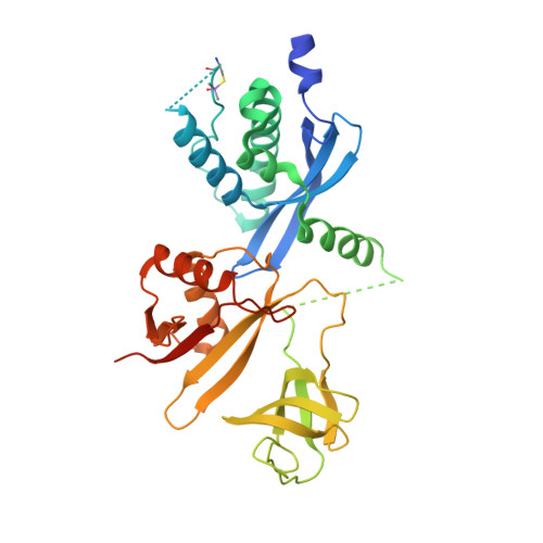
    The superoxide-producing phagocyte NADPH oxidase is activated during phagocytosis to destroy ingested microbes. The adaptor protein p40phox associates via the PB1 domain with the essential oxidase activator p67phox, and is considered to function by recruiting p67phox to phagosomes; in this process, the PX domain of p40phox binds to phosphatidylinositol 3-phosphate [PtdIns(3)P], a lipid abundant in the phagosomal membrane. Here we show that the PtdIns(3)P-binding activity of p40phox is normally inhibited by the PB1 domain both in vivo and in vitro. The crystal structure of the full-length p40phox reveals that the inhibition is mediated via intramolecular interaction between the PB1 and PX domains. The interface of the p40phox PB1 domain for the PX domain localizes on the opposite side of that for the p67phox PB1 domain, and thus the PB1-mediated PX regulation occurs without preventing the PB1-PB1 association with p67phox.


  • Organizational Affiliation

    Laboratory of Structural Biology, Graduate School of Pharmaceutical Sciences, Hokkaido University, Sapporo, Japan.


Macromolecules
Find similar proteins by:  (by identity cutoff)  |  3D Structure
Entity ID: 1
MoleculeChains Sequence LengthOrganismDetailsImage
Neutrophil cytosol factor 4
A, B
341Homo sapiensMutation(s): 0 
UniProt & NIH Common Fund Data Resources
Find proteins for Q15080 (Homo sapiens)
Explore Q15080 
Go to UniProtKB:  Q15080
PHAROS:  Q15080
GTEx:  ENSG00000100365 
Entity Groups  
Sequence Clusters30% Identity50% Identity70% Identity90% Identity95% Identity100% Identity
UniProt GroupQ15080
Sequence Annotations
Expand
  • Reference Sequence
Small Molecules
Modified Residues  1 Unique
IDChains TypeFormula2D DiagramParent
CAF
Query on CAF
A, B
L-PEPTIDE LINKINGC5 H12 As N O3 SCYS
Experimental Data & Validation

Experimental Data

  • Method: X-RAY DIFFRACTION
  • Resolution: 3.15 Å
  • R-Value Free: 0.302 
  • R-Value Work: 0.263 
  • R-Value Observed: 0.263 
  • Space Group: C 2 2 21
Unit Cell:
Length ( Å )Angle ( ˚ )
a = 146.273α = 90
b = 189.813β = 90
c = 79.883γ = 90
Software Package:
Software NamePurpose
CNSrefinement
HKL-2000data collection
HKL-2000data reduction
HKL-2000data scaling
MOLREPphasing

Structure Validation

View Full Validation Report


Entry History 

Deposition Data

  • Released Date: 2007-01-23 
  • Deposition Author(s): Honbou, K.

Revision History  (Full details and data files)

  • Version 1.0: 2007-01-23
    Type: Initial release
  • Version 1.1: 2008-04-30
    Changes: Version format compliance
  • Version 1.2: 2011-07-13
    Changes: Version format compliance
  • Version 1.3: 2023-10-25
    Changes: Data collection, Database references, Derived calculations, Refinement description