The structure and function of a novel glycerophosphodiesterase from Enterobacter aerogenes
Jackson, C.J., Carr, P.D., Liu, J.W., Watt, S.J., Beck, J.L., Ollis, D.L.(2007) J Mol Biol 367: 1047-1062
- PubMed: 17306828
- DOI: https://doi.org/10.1016/j.jmb.2007.01.032
- Primary Citation of Related Structures:
2DXL, 2DXN - PubMed Abstract:
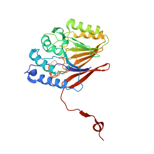
The structure of the glycerophosphodiesterase (GDPD) from Enterobacter aerogenes, GpdQ, has been solved by SAD phasing from the active site metal ions. Structural analysis indicates that GpdQ belongs to the alpha/beta sandwich metallo-phosphoesterase family, rather than the (alpha/beta)(8) barrel GDPD family, suggesting that GpdQ is a structurally novel GDPD. Hexameric GpdQ is generated by interactions between three dimers. The dimers are formed through domain swapping, stabilised by an inter-chain disulfide bond, and beta-sheet extension. The active site contains a binuclear metal centre, with a fully occupied alpha-metal ion site, and partially occupied beta-metal ion site, as revealed by anomalous scattering analysis. Using a combination of TLS refinement and normal mode analysis, the dynamic movement of GpdQ was investigated. This analysis suggests that the hexameric quaternary structure stabilises the base of the dimer, which promotes "breathing" of the active site cleft. Comparison with other metallo-phosphodiesterases shows that although the central, catalytic, domain is highly conserved, many of these enzymes possess structurally unrelated secondary domains located at the entrance of the active site. We suggest that this could be a common structural feature of metallo-phosphodiesterases that constrains substrate specificity, preventing non-specific phosphodiester hydrolysis.
Organizational Affiliation:
Research School of Chemistry, Australian National University, ACT, 0200, Australia.