Structure of aspartate racemase complexed with a dual substrate analogue, citric acid, and implications for the reaction mechanism.
Ohtaki, A., Nakano, Y., Iizuka, R., Arakawa, T., Yamada, K., Odaka, M., Yohda, M.(2008) Proteins 70: 1167-1174
- PubMed: 17847084
- DOI: https://doi.org/10.1002/prot.21528
- Primary Citation of Related Structures:
2DX7 - PubMed Abstract:
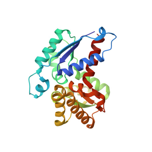
Pyrococcus horikoshii OT3 aspartate racemase (PhAspR) catalyzes the interconversion between L- and D-aspartate. The X-ray structure of PhAspR revealed a pseudo mirror-symmetric distribution of the residues around its active site, which is very reasonable for its chiral substrates, L-aspartate and D-aspartate. In this study, we have determined the crystal structure of an inactive mutant PhAspR complexed with a citric acid (Cit) at a resolution of 2.0 A. Cit contains the substrate analogue moieties of both L- and D-aspartate and exhibits a low competitive inhibition activity against PhAspR. In the structure, Cit binds to the catalytic site of PhAspR, which induced the conformational change to close the active site. The distance between the thiolates was estimated to be 7.4 A, representing a catalytic state and the substrate binding modes of PhAspR. Two conserved basic residues, Arg48 and Lys164, seem to be indispensable for PhAspR activity. Arg48 is thought to be responsible for recognizing carboxyl groups of the substrates L-/D-aspartates and stabilizing a reaction intermediate, and Lys164 is responsible for stabilizing a closed state structure. In this structure, the L-aspartate moiety of Cit is likely to take the substrate position of the PhAspR-substrate complex, which is very similar to that of Glutamate racemase. There is also another possibility that the two substrate analogue moieties of the bound Cit reflect the binding modes of both L- and D-aspartates. Based on the PhAspR-Cit complex structure, the reaction mechanism of aspartate racemase was elucidated.
Organizational Affiliation:
Department of Biotechnology and Life Science, Tokyo University of Agriculture & Technology, Koganei, Tokyo 184-8588, Japan.