Structural analysis of Bordetella pertussis BugE solute receptor in a bound conformation
Huvent, I., Belrhali, H., Antoine, R., Bompard, C., Locht, C., Jacob-Dubuisson, F., Villeret, V.(2006) Acta Crystallogr D Biol Crystallogr 62: 1375-1381
- PubMed: 17057341
- DOI: https://doi.org/10.1107/S0907444906032653
- Primary Citation of Related Structures:
2DVZ - PubMed Abstract:
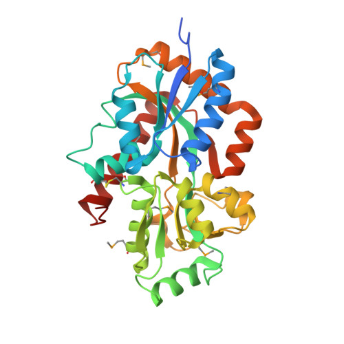
The Bug proteins form a large family of periplasmic solute-binding receptors present in a number of bacterial species. Here, the crystal structure of Bordetella pertussis BugE, a member of the Bug family coded by the gene BP0250, is reported. It adopts the Venus flytrap architecture of periplasmic binding proteins, with two domains separated by a deep cleft. BugE has a bound ligand, identified as a glutamate. The structure of B. pertussis BugD, which is an aspartic acid transporter, has recently been reported. These structures reveal high conservation of the Bug architecture, despite limited sequence identity. They share a common carboxylate-binding motif defined by two strand-beta-turn-alpha-helix motifs, also involving two water molecules to bridge the carboxylate O atoms to the protein. The two water molecules are hydrogen bonded to a common main-chain carbonyl group. Although the features of the carboxylate-binding motif are totally conserved, the ligand in BugE is bound by its side-chain carboxylate group rather than by its alpha-carboxylate as in BugD. This specific ligand-binding motif is highly conserved in Bug proteins and the BugE structure suggests that the cavity of the Bug proteins might also accommodate carboxylated solutes other than amino acids. The vast expansion of the Bug family in several bacterial genera is likely to be explained by the possible diversity of ligands. No charged residues are involved in glutamate binding by BugE, unlike what has been described for all glutamate receptors reported so far.
Organizational Affiliation:
UMR8161 CNRS, Institut de Biologie de Lille, Institute Pasteur de Lille, 1 rue du Professeur Calmette, BP245, 59021 Lille CEDEX, France.