2DU7

Crystal structure of Methanococcus jannacshii O-phosphoseryl-tRNA synthetase


Experimental Data Snapshot

  • Method: X-RAY DIFFRACTION
  • Resolution: 3.60 Å
  • R-Value Free: 0.387 
  • R-Value Work: 0.330 
  • R-Value Observed: 0.330 

wwPDB Validation   3D Report Full Report


This is version 1.3 of the entry. See complete history


Literature

Structural insights into the first step of RNA-dependent cysteine biosynthesis in archaea.

Fukunaga, R.Yokoyama, S.

(2007) Nat Struct Mol Biol 14: 272-279

  • DOI: https://doi.org/10.1038/nsmb1219
  • Primary Citation of Related Structures:  
    2DU3, 2DU4, 2DU5, 2DU6, 2DU7

  • PubMed Abstract: 

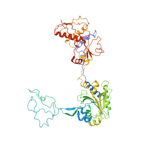
    Cysteine is ligated to tRNA(Cys) by cysteinyl-tRNA synthetase in most organisms. However, in methanogenic archaea lacking cysteinyl-tRNA synthetase, O-phosphoserine is ligated to tRNA(Cys) by O-phosphoseryl-tRNA synthetase (SepRS), and the phosphoseryl-tRNA(Cys) is converted to cysteinyl-tRNA(Cys). In this study, we determined the crystal structure of the SepRS tetramer in complex with tRNA(Cys) and O-phosphoserine at 2.6-A resolution. The catalytic domain of SepRS recognizes the negatively charged side chain of O-phosphoserine at a noncanonical site, using the dipole moment of a conserved alpha-helix. The unique C-terminal domain specifically recognizes the anticodon GCA of tRNA(Cys). On the basis of the structure, we engineered SepRS to recognize tRNA(Cys) mutants with the anticodons UCA and CUA and clarified the anticodon recognition mechanism by crystallography. The mutant SepRS-tRNA pairs may be useful for translational incorporation of O-phosphoserine into proteins in response to the stop codons UGA and UAG.


  • Organizational Affiliation

    Department of Biophysics and Biochemistry, Graduate School of Science, University of Tokyo, 7-3-1 Hongo, Tokyo 113-0033, Japan.


Macromolecules
Find similar proteins by:  (by identity cutoff)  |  3D Structure
Entity ID: 1
MoleculeChains Sequence LengthOrganismDetailsImage
O-phosphoseryl-tRNA synthetase
A, B, C, D
549Methanocaldococcus jannaschiiMutation(s): 0 
EC: 6.1.1
UniProt
Find proteins for Q59054 (Methanocaldococcus jannaschii (strain ATCC 43067 / DSM 2661 / JAL-1 / JCM 10045 / NBRC 100440))
Explore Q59054 
Go to UniProtKB:  Q59054
Entity Groups  
Sequence Clusters30% Identity50% Identity70% Identity90% Identity95% Identity100% Identity
UniProt GroupQ59054
Sequence Annotations
Expand
  • Reference Sequence
Experimental Data & Validation

Experimental Data

  • Method: X-RAY DIFFRACTION
  • Resolution: 3.60 Å
  • R-Value Free: 0.387 
  • R-Value Work: 0.330 
  • R-Value Observed: 0.330 
  • Space Group: C 2 2 21
Unit Cell:
Length ( Å )Angle ( ˚ )
a = 196.354α = 90
b = 299.455β = 90
c = 125.877γ = 90
Software Package:
Software NamePurpose
CNSrefinement
HKL-2000data reduction
HKL-2000data scaling
MOLREPphasing

Structure Validation

View Full Validation Report


Entry History 

Deposition Data

Revision History  (Full details and data files)

  • Version 1.0: 2007-03-13
    Type: Initial release
  • Version 1.1: 2007-10-04
    Changes: Version format compliance
  • Version 1.2: 2011-07-13
    Changes: Derived calculations, Version format compliance
  • Version 1.3: 2023-10-25
    Changes: Data collection, Database references, Refinement description