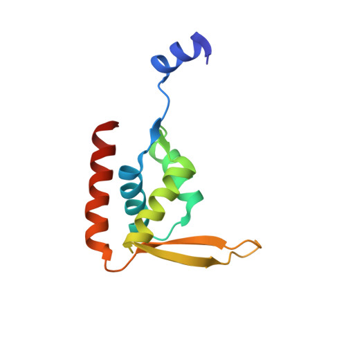
Functionally important structural elements of the cyanobacterial clock-related protein Pex.
Kurosawa, S., Murakami, R., Onai, K., Morishita, M., Hasegawa, D., Iwase, R., Uzumaki, T., Hayashi, F., Kitajima-Ihara, T., Sakata, S., Murakami, M., Kouyama, T., Ishiura, M.(2009) Genes Cells 14: 1-16
- PubMed: 19032344
- DOI: https://doi.org/10.1111/j.1365-2443.2008.01245.x
- Primary Citation of Related Structures:
2DQL, 2ZFW - PubMed Abstract:
Pex, a clock-related protein involved in the input pathway of the cyanobacterial circadian clock system, suppresses the expression of clock gene kaiA and lengthens the circadian period. Here, we determined the crystal structure of Anabaena Pex (AnaPex; Anabaena sp. strain PCC 7120) and Synechococcus Pex (SynPex; Synechococcus sp. strain PCC 7942). Pex is a homodimer that forms a winged-helix structure. Using the DNase I protection and electrophoresis mobility shift assays on a Synechococcus kaiA upstream region, we identified a minimal 25-bp sequence that contained an imperfectly inverted repeat sequence as the Pex-binding sequence. Based on crystal structure, we predicted the amino acid residues essential for Pex's DNA-binding activity and examined the effects of various Ala-substitutions in the alpha3 helix and wing region of Pex on in vitro DNA-binding activity and in vivo rhythm functions. Mutant AnaPex proteins carrying a substitution in the wing region displayed no specific DNA-binding activity, whereas those carrying a substitution in the alpha3 helix did display specific binding activity. But the latter were less thermostable than wild-type AnaPex and their in vitro functions were defective. We concluded that Pex binds a kaiA upstream DNA sequence via its wing region and that its alpha3 helix is probably important to its stability.
Organizational Affiliation:
Department of Physics, Graduate School of Science, Nagoya University, Furo, Chikusa, Nagoya 464-8602, Japan.