2DOK

Crystal structure of the PIN domain of human EST1A


Experimental Data Snapshot

  • Method: X-RAY DIFFRACTION
  • Resolution: 1.80 Å
  • R-Value Free: 0.254 
  • R-Value Work: 0.213 
  • R-Value Observed: 0.215 

wwPDB Validation   3D Report Full Report


This is version 1.3 of the entry. See complete history


Literature

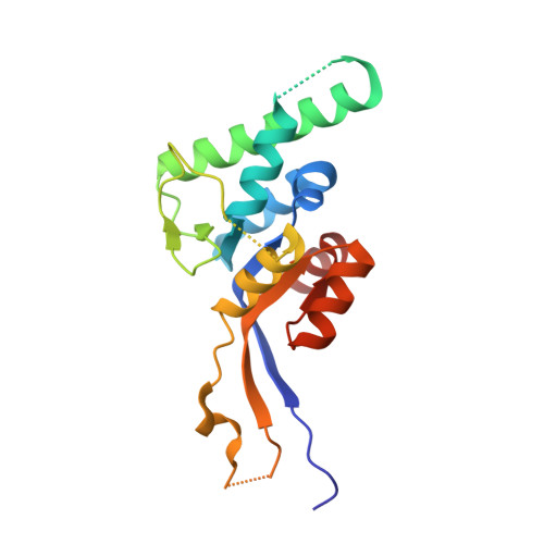
Crystal structure of the PIN domain of human telomerase-associated protein EST1A

Takeshita, D.Zenno, S.Lee, W.C.Saigo, K.Tanokura, M.

(2007) Proteins 68: 980-989

  • DOI: https://doi.org/10.1002/prot.21351
  • Primary Citation of Related Structures:  
    2DOK

  • PubMed Abstract: 

    Saccharomyces cerevisiae Est1p is a telomerase-associated protein essential for telomere length homeostasis. hEST1A is one of the three human Est1p homologues and is considered to be involved not only in regulation of telomere elongation or capping but also in nonsense-mediated degradation of RNA. hEST1A is composed of two conserved regions, Est1p homology and PIN (PilT N-terminus) domains. The present study shows the crystal structure of the PIN domain at 1.8 A resolution. The overall structure is composed of an alpha/beta fold or a core structure similar to the counterpart of 5' nucleases and an extended structure absent from archaeal PIN-domain proteins and 5' nucleases. The structural properties of the PIN domain indicate its putative active center consisting of invariant acidic amino acid residues, which is geometrically similar to the active center of 5' nucleases and an archaeal PAE2754 PIN-domain protein associated with exonuclease activity.


  • Organizational Affiliation

    Department of Applied Biological Chemistry, Graduate School of Agricultural and Life Sciences, University of Tokyo, Bunkyo-ku, Tokyo 113-8657, Japan.


Macromolecules
Find similar proteins by:  (by identity cutoff)  |  3D Structure
Entity ID: 1
MoleculeChains Sequence LengthOrganismDetailsImage
Telomerase-binding protein EST1A
A, B
186Homo sapiensMutation(s): 0 
Gene Names: EST1A
UniProt & NIH Common Fund Data Resources
Find proteins for Q86US8 (Homo sapiens)
Explore Q86US8 
Go to UniProtKB:  Q86US8
PHAROS:  Q86US8
GTEx:  ENSG00000070366 
Entity Groups  
Sequence Clusters30% Identity50% Identity70% Identity90% Identity95% Identity100% Identity
UniProt GroupQ86US8
Sequence Annotations
Expand
  • Reference Sequence
Experimental Data & Validation

Experimental Data

  • Method: X-RAY DIFFRACTION
  • Resolution: 1.80 Å
  • R-Value Free: 0.254 
  • R-Value Work: 0.213 
  • R-Value Observed: 0.215 
  • Space Group: C 1 2 1
Unit Cell:
Length ( Å )Angle ( ˚ )
a = 107.326α = 90
b = 51.586β = 119.25
c = 100.48γ = 90
Software Package:
Software NamePurpose
REFMACrefinement
HKL-2000data reduction
SCALEPACKdata scaling
CNSphasing

Structure Validation

View Full Validation Report


Entry History 

Deposition Data

Revision History  (Full details and data files)

  • Version 1.0: 2007-05-01
    Type: Initial release
  • Version 1.1: 2007-10-28
    Changes: Version format compliance
  • Version 1.2: 2011-07-13
    Changes: Derived calculations, Version format compliance
  • Version 1.3: 2024-03-13
    Changes: Data collection, Database references