2DNJ

DNASE I-INDUCED DNA CONFORMATION. 2 ANGSTROMS STRUCTURE OF A DNASE I-OCTAMER COMPLEX


Experimental Data Snapshot

  • Method: X-RAY DIFFRACTION
  • Resolution: 2.00 Å
  • R-Value Observed: 0.174 

wwPDB Validation   3D Report Full Report


This is version 2.0 of the entry. See complete history


Literature

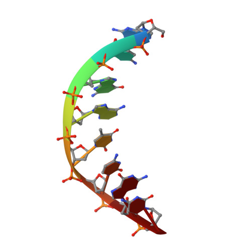
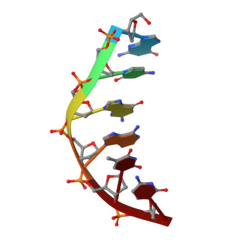
DNase I-induced DNA conformation. 2 A structure of a DNase I-octamer complex

Lahm, A.Suck, D.

(1991) J Mol Biol 222: 645-667

  • DOI: https://doi.org/10.1016/0022-2836(91)90502-w
  • Primary Citation of Related Structures:  
    2DNJ

  • PubMed Abstract: 

    The structure of a complex between DNase I and d(GCGATCGC)2 has been solved by molecular replacement and refined to an R-factor of 0.174 for all data between 6 and 2 A resolution. The nicked octamer duplexes have lost a dinucleotide from the 3' ends of one strand and are hydrogen-bonded across a 2-fold axis to form a quasi-continuous double helix of 14 base-pairs. DNase I is bound in the minor groove of the B-type DNA duplex forming contacts in and along both sides of the minor groove extending over a total of six base-pairs. As a consequence of binding of DNase I to the DNA-substrate the minor groove opens by about 3 A and the duplex bends towards the major groove by about 20 degrees. Apart from these more global distortions the bound duplex also shows significant deviations in local geometry. A major cause for the observed perturbations in the DNA conformation seems to be the stacking type interaction of a tyrosine ring (Y76) with a deoxyribose. In contrast, the enzyme structure is nearly unchanged compared to free DNase I (0.49 A root-mean-square deviations for main-chain atoms) thus providing a rigid framework to which the DNA substrate has to adapt on binding. These results confirm the hypothesis that groove width and stiffness are major factors determining the global sequence dependence of the enzyme's cutting rates. The nicked octamer present in the crystals did not allow us to draw detailed conclusions about the catalytic mechanism but confirmed the location of the active site near H134 on top of the central beta-sheets. A second cut of the DNA induced by diffusion of Mn2+ into the crystals may suggest the presence of a secondary active site in DNase I.


  • Organizational Affiliation

    European Molecular Biology Laboratory, Biological Structures Division, Heidelberg, Germany.


Macromolecules

Find similar proteins by:  (by identity cutoff)  |  3D Structure
Entity ID: 3
MoleculeChains Sequence LengthOrganismDetailsImage
DEOXYRIBONUCLEASE IC [auth A]260Bos taurusMutation(s): 0 
EC: 3.1.21.1
UniProt
Find proteins for P00639 (Bos taurus)
Explore P00639 
Go to UniProtKB:  P00639
Entity Groups  
Sequence Clusters30% Identity50% Identity70% Identity90% Identity95% Identity100% Identity
UniProt GroupP00639
Sequence Annotations
Expand
  • Reference Sequence

Find similar nucleic acids by:  Sequence   |   3D Structure  

Entity ID: 1
MoleculeChains LengthOrganismImage
5'-D(*GP*CP*GP*AP*TP*CP*GP*C)-3'A [auth B]8N/A
Sequence Annotations
Expand
  • Reference Sequence

Find similar nucleic acids by:  Sequence   |   3D Structure  

Entity ID: 2
MoleculeChains LengthOrganismImage
5'-D(*GP*CP*GP*AP*TP*C)-3'B [auth C]6N/A
Sequence Annotations
Expand
  • Reference Sequence
Small Molecules
Ligands 1 Unique
IDChains Name / Formula / InChI Key2D Diagram3D Interactions
NAG
Query on NAG

Download Ideal Coordinates CCD File 
D [auth A]2-acetamido-2-deoxy-beta-D-glucopyranose
C8 H15 N O6
OVRNDRQMDRJTHS-FMDGEEDCSA-N
Experimental Data & Validation

Experimental Data

  • Method: X-RAY DIFFRACTION
  • Resolution: 2.00 Å
  • R-Value Observed: 0.174 
  • Space Group: C 2 2 21
Unit Cell:
Length ( Å )Angle ( ˚ )
a = 72.9α = 90
b = 100.1β = 90
c = 92.6γ = 90
Software Package:
Software NamePurpose
PROLSQrefinement

Structure Validation

View Full Validation Report


Entry History 

Deposition Data

Revision History  (Full details and data files)

  • Version 1.0: 1994-01-31
    Type: Initial release
  • Version 1.1: 2008-05-22
    Changes: Version format compliance
  • Version 1.2: 2011-07-13
    Changes: Version format compliance
  • Version 1.3: 2017-11-29
    Changes: Derived calculations
  • Version 1.4: 2020-07-29
    Type: Remediation
    Reason: Carbohydrate remediation
    Changes: Data collection, Derived calculations, Structure summary
  • Version 2.0: 2020-10-21
    Changes: Advisory, Atomic model, Data collection, Derived calculations, Non-polymer description, Structure summary