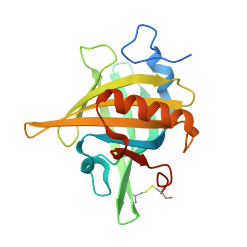
2DM5

Thermodynamic Penalty Arising From Burial of a Ligand Polar Group Within a Hydrophobic Pocket of a Protein Receptor


Experimental Data Snapshot

  • Method: X-RAY DIFFRACTION
  • Resolution: 1.70 Å
  • R-Value Free: 0.200 
  • R-Value Work: 0.190 
  • R-Value Observed: 0.190 

wwPDB Validation   3D Report Full Report


This is version 1.3 of the entry. See complete history


Literature

Thermodynamic penalty arising from burial of a ligand polar group within a hydrophobic pocket of a protein receptor

Barratt, E.Bronowska, A.Vondrasek, J.Cerny, J.Bingham, R.Phillips, S.Homans, S.W.

(2006) J Mol Biol 362: 994-1003

  • DOI: https://doi.org/10.1016/j.jmb.2006.07.067
  • Primary Citation of Related Structures:  
    2DM5

  • PubMed Abstract: 

    Here, we examine the thermodynamic penalty arising from burial of a polar group in a hydrophobic pocket that forms part of the binding-site of the major urinary protein (MUP-I). X-ray crystal structures of the complexes of octanol, nonanol and 1,8 octan-diol indicate that these ligands bind with similar orientations in the binding pocket. Each complex is characterised by a bridging water molecule between the hydroxyl group of Tyr120 and the hydroxyl group of each ligand. The additional hydroxyl group of 1,8 octan-diol is thereby forced to reside in a hydrophobic pocket, and isothermal titration calorimetry experiments indicate that this is accompanied by a standard free energy penalty of +21 kJ/mol with respect to octanol and +18 kJ/mol with respect to nonanol. Consideration of the solvation thermodynamics of each ligand enables the "intrinsic" (solute-solute) interaction energy to be determined, which indicates a favourable enthalpic component and an entropic component that is small or zero. These data indicate that the thermodynamic penalty to binding derived from the unfavourable desolvation of 1,8 octan-diol is partially offset by a favourable intrinsic contribution. Quantum chemical calculations suggest that this latter contribution derives from favourable solute-solute dispersion interactions.


  • Organizational Affiliation

    Astbury Centre for Structural Molecular Biology, School of Biochemistry and Molecular Biology, University of Leeds, LS2 9JT, UK.


Macromolecules
Find similar proteins by:  (by identity cutoff)  |  3D Structure
Entity ID: 1
MoleculeChains Sequence LengthOrganismDetailsImage
Major Urinary Protein174Mus musculusMutation(s): 0 
Gene Names: MUP1
UniProt
Find proteins for P11589 (Mus musculus)
Explore P11589 
Go to UniProtKB:  P11589
Entity Groups  
Sequence Clusters30% Identity50% Identity70% Identity90% Identity95% Identity100% Identity
UniProt GroupP11589
Sequence Annotations
Expand
  • Reference Sequence
Small Molecules
Binding Affinity Annotations 
IDSourceBinding Affinity
ODI Binding MOAD:  2DM5 Kd: 1.01e+6 (nM) from 1 assay(s)
BindingDB:  2DM5 -TΔS: min: 10.66, max: 17.91 (kJ/mol) from 3 assay(s)
ΔG: min: -3.39e+1, max: -1.72e+1 (kJ/mol) from 3 assay(s)
Experimental Data & Validation

Experimental Data

  • Method: X-RAY DIFFRACTION
  • Resolution: 1.70 Å
  • R-Value Free: 0.200 
  • R-Value Work: 0.190 
  • R-Value Observed: 0.190 
  • Space Group: P 43 21 2
Unit Cell:
Length ( Å )Angle ( ˚ )
a = 53.522α = 90
b = 53.522β = 90
c = 137.192γ = 90
Software Package:
Software NamePurpose
CrystalCleardata collection
SCALAdata scaling
CNSrefinement
CrystalCleardata reduction
CCP4data scaling
CNSphasing

Structure Validation

View Full Validation Report


Entry History 

Deposition Data

Revision History  (Full details and data files)

  • Version 1.0: 2006-10-17
    Type: Initial release
  • Version 1.1: 2008-04-30
    Changes: Version format compliance
  • Version 1.2: 2011-07-13
    Changes: Version format compliance
  • Version 1.3: 2023-10-25
    Changes: Data collection, Database references, Derived calculations, Refinement description