2DGJ

Crystal structure of EbhA (756-1003 domain) from Staphylococcus aureus


Experimental Data Snapshot

  • Method: X-RAY DIFFRACTION
  • Resolution: 2.35 Å
  • R-Value Free: 0.287 
  • R-Value Work: 0.241 
  • R-Value Observed: 0.241 

wwPDB Validation   3D Report Full Report


This is version 1.2 of the entry. See complete history


Literature

A helical string of alternately connected three-helix bundles for the cell wall-associated adhesion protein Ebh from Staphylococcus aureus

Tanaka, Y.Sakamoto, S.Kuroda, M.Goda, S.Gao, Y.-G.Tsumoto, K.Hiragi, Y.Yao, M.Watanabe, N.Ohta, T.Tanaka, I.

(2008) Structure 16: 488-496

  • DOI: https://doi.org/10.1016/j.str.2007.12.018
  • Primary Citation of Related Structures:  
    2DGJ

  • PubMed Abstract: 

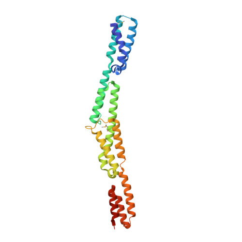
    The 1.1 MDa cell-wall-associated adhesion protein of staphylococci, Ebh, consists of several distinct regions, including a large central region with 52 imperfect repeats of 126 amino acid residues. We investigated the structure of this giant molecule by X-ray crystallography, circular dichroism (CD) spectrometry, and small-angle X-ray scattering (SAXS). The crystal structure of two repeats showed that each repeat consists of two distinct three-helix bundles, and two such repeats are connected along the long axis, resulting in a rod-like structure that is 120 A in length. CD and SAXS analyses of the samples with longer repeats suggested that each repeat has an identical structure, and that such repeats are connected tandemly to form a rod-like structure in solution, the length of which increased proportionately with the number of repeating units. On the basis of these results, it was proposed that Ebh is a 320 nm rod-like molecule with some plasticity at module junctions.


  • Organizational Affiliation

    Faculty of Advanced Life Sciences, Hokkaido University, Sapporo 060-0810, Japan.


Macromolecules
Find similar proteins by:  (by identity cutoff)  |  3D Structure
Entity ID: 1
MoleculeChains Sequence LengthOrganismDetailsImage
hypothetical protein ebhA
A, B
257Staphylococcus aureusMutation(s): 0 
UniProt
Find proteins for Q931R6 (Staphylococcus aureus (strain Mu50 / ATCC 700699))
Explore Q931R6 
Go to UniProtKB:  Q931R6
Entity Groups  
Sequence Clusters30% Identity50% Identity70% Identity90% Identity95% Identity100% Identity
UniProt GroupQ931R6
Sequence Annotations
Expand
  • Reference Sequence
Experimental Data & Validation

Experimental Data

  • Method: X-RAY DIFFRACTION
  • Resolution: 2.35 Å
  • R-Value Free: 0.287 
  • R-Value Work: 0.241 
  • R-Value Observed: 0.241 
  • Space Group: C 2 2 21
Unit Cell:
Length ( Å )Angle ( ˚ )
a = 45.265α = 90
b = 161.848β = 90
c = 133.366γ = 90
Software Package:
Software NamePurpose
CNSrefinement
HKL-2000data reduction
HKL-2000data scaling
SHELXphasing
OASISphasing
DMphasing
SOLVEphasing

Structure Validation

View Full Validation Report


Entry History 

Deposition Data

Revision History  (Full details and data files)

  • Version 1.0: 2007-03-20
    Type: Initial release
  • Version 1.1: 2008-04-30
    Changes: Version format compliance
  • Version 1.2: 2011-07-13
    Changes: Non-polymer description, Version format compliance