Three-dimensional structure of the myosin V inhibited state by cryoelectron tomography
Liu, J., Taylor, D.W., Krementsova, E.B., Trybus, K.M., Taylor, K.A.(2006) Nature 442: 208-211
- PubMed: 16625208
- DOI: https://doi.org/10.1038/nature04719
- Primary Citation of Related Structures:
2DFS - PubMed Abstract:
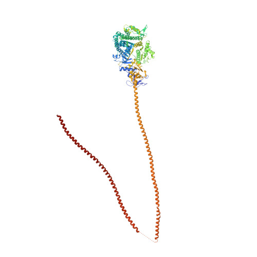
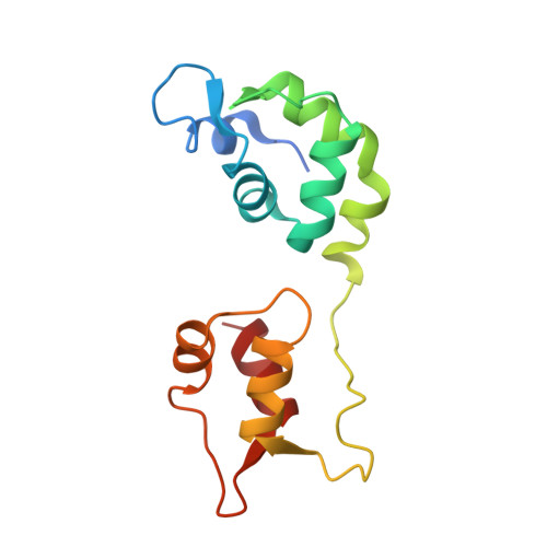
Unconventional myosin V (myoV) is an actin-based molecular motor that has a key function in organelle and mRNA transport, as well as in membrane trafficking. MyoV was the first member of the myosin superfamily shown to be processive, meaning that a single motor protein can 'walk' hand-over-hand along an actin filament for many steps before detaching. Full-length myoV has a low actin-activated MgATPase activity at low [Ca2+], whereas expressed constructs lacking the cargo-binding domain have a high activity regardless of [Ca2+] (refs 5-7). Hydrodynamic data and electron micrographs indicate that the active state is extended, whereas the inactive state is compact. Here we show the first three-dimensional structure of the myoV inactive state. Each myoV molecule consists of two heads that contain an amino-terminal motor domain followed by a lever arm that binds six calmodulins. The heads are followed by a coiled-coil dimerization domain (S2) and a carboxy-terminal globular cargo-binding domain. In the inactive structure, bending of myoV at the head-S2 junction places the cargo-binding domain near the motor domain's ATP-binding pocket, indicating that ATPase inhibition might occur through decreased rates of nucleotide exchange. The actin-binding interfaces are unobstructed, and the lever arm is oriented in a position typical of strong actin-binding states. This structure indicates that motor recycling after cargo delivery might occur through transport on actively treadmilling actin filaments rather than by diffusion.
Organizational Affiliation:
The Institute of Molecular Biophysics, Florida State University, Tallahassee, Florida 32306-4380, USA.