Automated protein structure determination from NMR spectra
Lopez-Mendez, B., Guntert, P.(2006) J Am Chem Soc 128: 13112-13122
- PubMed: 17017791
- DOI: https://doi.org/10.1021/ja061136l
- Primary Citation of Related Structures:
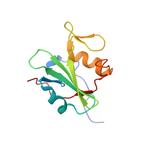
2DCP, 2DCQ, 2DCR - PubMed Abstract:
Fully automated structure determination of proteins in solution (FLYA) yields, without human intervention, three-dimensional protein structures starting from a set of multidimensional NMR spectra. Integrating existing and new software, automated peak picking over all spectra is followed by peak list filtering, the generation of an ensemble of initial chemical shift assignments, the determination of consensus chemical shift assignments for all (1)H, (13)C, and (15)N nuclei, the assignment of NOESY cross-peaks, the generation of distance restraints, and the calculation of the three-dimensional structure by torsion angle dynamics. The resulting, preliminary structure serves as additional input to the second stage of the procedure, in which a new ensemble of chemical shift assignments and a refined structure are calculated. The three-dimensional structures of three 12-16 kDa proteins computed with the FLYA algorithm coincided closely with the conventionally determined structures. Deviations were below 0.95 A for the backbone atom positions, excluding the flexible chain termini. 96-97% of all backbone and side-chain chemical shifts in the structured regions were assigned to the correct residues. The purely computational FLYA method is suitable for substituting all manual spectra analysis and thus overcomes a main efficiency limitation of the NMR method for protein structure determination.
Organizational Affiliation:
Tatsuo Miyazawa Memorial Program, RIKEN Genomic Sciences Center, Tsurumi, Yokohama 230-0045, Japan.