2DCF

Crystal structure of 6-aminohexanoate-dimer hydrolase S112A/G181D/H266N mutant with substrate


Experimental Data Snapshot

  • Method: X-RAY DIFFRACTION
  • Resolution: 1.40 Å
  • R-Value Free: 0.195 
  • R-Value Work: 0.183 
  • R-Value Observed: 0.183 

wwPDB Validation   3D Report Full Report


This is version 2.0 of the entry. See complete history


Literature

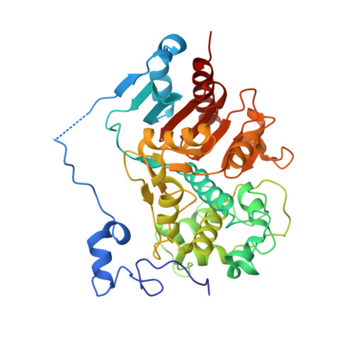
Nylon-oligomer degrading enzyme/substrate complex: catalytic mechanism of 6-aminohexanoate-dimer hydrolase

Negoro, S.Ohki, T.Shibata, N.Sasa, K.Hayashi, H.Nakano, H.Yasuhira, K.Kato, D.Takeo, M.Higuchi, Y.

(2007) J Mol Biol 370: 142-156

  • DOI: https://doi.org/10.1016/j.jmb.2007.04.043
  • Primary Citation of Related Structures:  
    1WYC, 2DCF

  • PubMed Abstract: 

    We performed X-ray crystallographic analyses of 6-aminohexanoate-dimer hydrolase (Hyb-24DN), an enzyme responsible for the degradation of nylon-6, an industry by-product, and of a complex between Hyb-24DN-A(112) (S112A-mutant of Hyb-24DN) and 6-aminohexanoate-linear dimer (Ald) at 1.58 A and 1.4 A resolution, respectively. In Hyb-24DN, Asp181-O(delta) forms hydrogen bonds with Tyr170-O(eta), -two of the catalytic and binding amino acids, and a loop between Asn167 and Val177. This state is the so-called open form, allowing its substrate to bind in the space between the loop and catalytic residues. Upon substrate binding (in Hyb-24DN-A(112)/Ald complex), the loop is shifted 4.3 A at Tyr170-C(alpha), and the side-chain of Tyr170 is rotated. By the combined effect, Tyr170-O(eta) moves a total of 10.5 A, resulting in the formation of hydrogen bonds with the nitrogen of amide linkage in Ald (closed form). In addition, electrostatic interaction between Asp181-O(delta) and the amino group in Ald stabilizes the substrate binding. We propose here that the enzyme catalysis proceeds according to the following steps: (i) Ald-induced transition from open to closed form, (ii) nucleophilic attack of Ser112 to Ald and formation of a tetrahedral intermediate, (iii) formation of acyl enzyme and transition to open form, (iv) deacylation. Amino acid substitutions reducing the enzyme/Ald interaction at positions 181 or 170 drastically decreased the Ald-hydrolytic activity, but had very little effect on esterolytic activity, suggesting that esterolytic reaction proceeds regardless of conversion. Present models illustrate why new activity against the nylon oligomer has evolved in an esterase with beta-lactamase folds, while retaining the original esterolytic functions.


  • Organizational Affiliation

    Department of Materials Science and Chemistry, Graduate School of Engineering, University of Hyogo, 2167 Shosha, Himeji, Hyogo 671-2280, Japan. negoro@eng.u-hyogo.ac.jp


Macromolecules
Find similar proteins by:  (by identity cutoff)  |  3D Structure
Entity ID: 1
MoleculeChains Sequence LengthOrganismDetailsImage
6-aminohexanoate-dimer hydrolase392Flavobacterium sp.Mutation(s): 3 
EC: 3.5.1.46
UniProt
Find proteins for P07062 (Paenarthrobacter ureafaciens)
Explore P07062 
Go to UniProtKB:  P07062
Entity Groups  
Sequence Clusters30% Identity50% Identity70% Identity90% Identity95% Identity100% Identity
UniProt GroupP07062
Sequence Annotations
Expand
  • Reference Sequence
Small Molecules
Ligands 4 Unique
IDChains Name / Formula / InChI Key2D Diagram3D Interactions
MES
Query on MES

Download Ideal Coordinates CCD File 
F [auth A]2-(N-MORPHOLINO)-ETHANESULFONIC ACID
C6 H13 N O4 S
SXGZJKUKBWWHRA-UHFFFAOYSA-N
ACA
Query on ACA

Download Ideal Coordinates CCD File 
D [auth A],
E [auth A]
6-AMINOHEXANOIC ACID
C6 H13 N O2
SLXKOJJOQWFEFD-UHFFFAOYSA-N
SO4
Query on SO4

Download Ideal Coordinates CCD File 
B [auth A],
C [auth A]
SULFATE ION
O4 S
QAOWNCQODCNURD-UHFFFAOYSA-L
GOL
Query on GOL

Download Ideal Coordinates CCD File 
G [auth A],
H [auth A],
I [auth A],
J [auth A]
GLYCEROL
C3 H8 O3
PEDCQBHIVMGVHV-UHFFFAOYSA-N
Experimental Data & Validation

Experimental Data

  • Method: X-RAY DIFFRACTION
  • Resolution: 1.40 Å
  • R-Value Free: 0.195 
  • R-Value Work: 0.183 
  • R-Value Observed: 0.183 
  • Space Group: P 32 2 1
Unit Cell:
Length ( Å )Angle ( ˚ )
a = 96.62α = 90
b = 96.62β = 90
c = 112.6γ = 120
Software Package:
Software NamePurpose
CNSrefinement
HKL-2000data reduction
SCALEPACKdata scaling
CNSphasing

Structure Validation

View Full Validation Report


Entry History 

Deposition Data

Revision History  (Full details and data files)

  • Version 1.0: 2007-01-09
    Type: Initial release
  • Version 1.1: 2008-04-30
    Changes: Version format compliance
  • Version 1.2: 2011-07-13
    Changes: Non-polymer description, Version format compliance
  • Version 1.3: 2017-08-23
    Changes: Source and taxonomy
  • Version 1.4: 2021-11-10
    Changes: Database references, Derived calculations
  • Version 1.5: 2023-10-25
    Changes: Data collection, Refinement description
  • Version 2.0: 2023-11-15
    Changes: Advisory, Atomic model, Data collection, Derived calculations