2DC1

Crystal Structure Of L-Aspartate Dehydrogenase From Hyperthermophilic Archaeon Archaeoglobus fulgidus


Experimental Data Snapshot

  • Method: X-RAY DIFFRACTION
  • Resolution: 1.90 Å
  • R-Value Free: 0.226 
  • R-Value Work: 0.217 

wwPDB Validation   3D Report Full Report


This is version 1.3 of the entry. See complete history


Literature

Crystal structure of archaeal highly thermostable L-aspartate dehydrogenase/NAD/citrate ternary complex.

Yoneda, K.Sakuraba, H.Tsuge, H.Katunuma, N.Ohshima, T.

(2007) FEBS J 274: 4315-4325

  • DOI: https://doi.org/10.1111/j.1742-4658.2007.05961.x
  • Primary Citation of Related Structures:  
    2DC1

  • PubMed Abstract: 

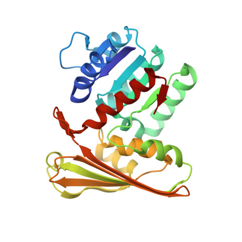
    The crystal structure of the highly thermostable L-aspartate dehydrogenase (L-aspDH; EC 1.4.1.21) from the hyperthermophilic archaeon Archaeoglobus fulgidus was determined in the presence of NAD and a substrate analog, citrate. The dimeric structure of A. fulgidus L-aspDH was refined at a resolution of 1.9 A with a crystallographic R-factor of 21.7% (R(free) = 22.6%). The structure indicates that each subunit consists of two domains separated by a deep cleft containing an active site. Structural comparison of the A. fulgidus L-aspDH/NAD/citrate ternary complex and the Thermotoga maritima L-aspDH/NAD binary complex showed that A. fulgidus L-aspDH assumes a closed conformation and that a large movement of the two loops takes place during substrate binding. Like T. maritima L-aspDH, the A. fulgidus enzyme is highly thermostable. But whereas a large number of inter- and intrasubunit ion pairs are responsible for the stability of A. fulgidus L-aspDH, a large number of inter- and intrasubunit aromatic pairs stabilize the T. maritima enzyme. Thus stabilization of these two L-aspDHs appears to be achieved in different ways. This is the first detailed description of substrate and coenzyme binding to L-aspDH and of the molecular basis of the high thermostability of a hyperthermophilic L-aspDH.


  • Organizational Affiliation

    Institute of Genetic Resources, Faculty of Agriculture, Kyushu University, Fukuoka, Japan.


Macromolecules
Find similar proteins by:  (by identity cutoff)  |  3D Structure
Entity ID: 1
MoleculeChains Sequence LengthOrganismDetailsImage
L-aspartate dehydrogenase
A, B
236Archaeoglobus fulgidusMutation(s): 0 
EC: 1.4.1.21
UniProt
Find proteins for O28440 (Archaeoglobus fulgidus (strain ATCC 49558 / DSM 4304 / JCM 9628 / NBRC 100126 / VC-16))
Explore O28440 
Go to UniProtKB:  O28440
Entity Groups  
Sequence Clusters30% Identity50% Identity70% Identity90% Identity95% Identity100% Identity
UniProt GroupO28440
Sequence Annotations
Expand
  • Reference Sequence
Small Molecules
Ligands 2 Unique
IDChains Name / Formula / InChI Key2D Diagram3D Interactions
NAD
Query on NAD

Download Ideal Coordinates CCD File 
D [auth A],
F [auth B]
NICOTINAMIDE-ADENINE-DINUCLEOTIDE
C21 H27 N7 O14 P2
BAWFJGJZGIEFAR-NNYOXOHSSA-N
CIT
Query on CIT

Download Ideal Coordinates CCD File 
C [auth A],
E [auth B]
CITRIC ACID
C6 H8 O7
KRKNYBCHXYNGOX-UHFFFAOYSA-N
Experimental Data & Validation

Experimental Data

  • Method: X-RAY DIFFRACTION
  • Resolution: 1.90 Å
  • R-Value Free: 0.226 
  • R-Value Work: 0.217 
  • Space Group: P 21 21 2
Unit Cell:
Length ( Å )Angle ( ˚ )
a = 47.52α = 90
b = 89.58β = 90
c = 100.49γ = 90
Software Package:
Software NamePurpose
HKL-2000data collection
SCALEPACKdata scaling
SOLVEphasing
CNSrefinement
HKL-2000data reduction

Structure Validation

View Full Validation Report


Entry History 

Deposition Data

Revision History  (Full details and data files)

  • Version 1.0: 2006-12-26
    Type: Initial release
  • Version 1.1: 2008-04-30
    Changes: Version format compliance
  • Version 1.2: 2011-07-13
    Changes: Version format compliance
  • Version 1.3: 2024-03-13
    Changes: Data collection, Database references, Derived calculations