2D4Z

Crystal structure of the cytoplasmic domain of the chloride channel ClC-0


Experimental Data Snapshot

  • Method: X-RAY DIFFRACTION
  • Resolution: 3.10 Å
  • R-Value Free: 0.309 
  • R-Value Work: 0.274 
  • R-Value Observed: 0.274 

wwPDB Validation   3D Report Full Report


This is version 1.3 of the entry. See complete history


Literature

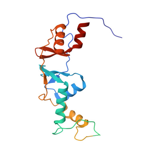
Crystal structure of the cytoplasmic domain of the chloride channel ClC-0.

Meyer, S.Dutzler, R.

(2006) Structure 14: 299-307

  • DOI: https://doi.org/10.1016/j.str.2005.10.008
  • Primary Citation of Related Structures:  
    2D4Z

  • PubMed Abstract: 

    Ion channels are frequently organized in a modular fashion and consist of a membrane-embedded pore domain and a soluble regulatory domain. A similar organization is found for the ClC family of Cl- channels and transporters. Here, we describe the crystal structure of the cytoplasmic domain of ClC-0, the voltage-dependent Cl- channel from T. marmorata. The structure contains a folded core of two tightly interacting cystathionine beta-synthetase (CBS) subdomains. The two subdomains are connected by a 96 residue mobile linker that is disordered in the crystals. As revealed by analytical ultracentrifugation, the domains form dimers, thereby most likely extending the 2-fold symmetry of the transmembrane pore. The structure provides insight into the organization of the cytoplasmic domains within the ClC family and establishes a framework for guiding future investigations on regulatory mechanisms.


  • Organizational Affiliation

    Department of Biochemistry, University of Zürich, Winterthurer Strasse 190, CH-8057 Zürich, Switzerland.


Macromolecules
Find similar proteins by:  (by identity cutoff)  |  3D Structure
Entity ID: 1
MoleculeChains Sequence LengthOrganismDetailsImage
Chloride channel protein
A, B
250Torpedo marmorataMutation(s): 0 
UniProt
Find proteins for P21564 (Torpedo marmorata)
Explore P21564 
Go to UniProtKB:  P21564
Entity Groups  
Sequence Clusters30% Identity50% Identity70% Identity90% Identity95% Identity100% Identity
UniProt GroupP21564
Sequence Annotations
Expand
  • Reference Sequence
Experimental Data & Validation

Experimental Data

  • Method: X-RAY DIFFRACTION
  • Resolution: 3.10 Å
  • R-Value Free: 0.309 
  • R-Value Work: 0.274 
  • R-Value Observed: 0.274 
  • Space Group: P 21 3
Unit Cell:
Length ( Å )Angle ( ˚ )
a = 125.819α = 90
b = 125.819β = 90
c = 125.819γ = 90
Software Package:
Software NamePurpose
CNSrefinement
HKL-2000data reduction
SCALEPACKdata scaling
SHARPphasing

Structure Validation

View Full Validation Report


Entry History 

Deposition Data

Revision History  (Full details and data files)

  • Version 1.0: 2006-02-14
    Type: Initial release
  • Version 1.1: 2008-04-30
    Changes: Version format compliance
  • Version 1.2: 2011-07-13
    Changes: Derived calculations, Version format compliance
  • Version 1.3: 2024-03-13
    Changes: Data collection, Database references