Refolding, characterization and crystal structure of (S)-malate dehydrogenase from the hyperthermophilic archaeon Aeropyrum pernix.
Kawakami, R., Sakuraba, H., Goda, S., Tsuge, H., Ohshima, T.(2009) Biochim Biophys Acta 1794: 1496-1504
- PubMed: 19555779
- DOI: https://doi.org/10.1016/j.bbapap.2009.06.014
- Primary Citation of Related Structures:
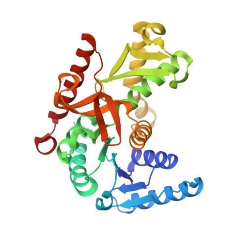
2D4A - PubMed Abstract:
Tartrate oxidation activity was found in the crude extract of an aerobic hyperthermophilic archaeon Aeropyrum pernix, and the enzyme was identified as (S)-malate dehydrogenase (MDH), which, when produced in Escherichia coli, was mainly obtained as an inactive inclusion body. The inclusion body was dissolved in 6 M guanidine-HCl and gradually refolded to the active enzyme through dilution of the denaturant. The purified recombinant enzyme consisted of four identical subunits with a molecular mass of about 110 kDa. NADP was preferred as a coenzyme over NAD for (S)-malate oxidation and, unlike MDHs from other sources, this enzyme readily catalyzed the oxidation of (2S,3S)-tartrate and (2S,3R)-tartrate. The tartrate oxidation activity was also observed in MDHs from the hyperthermophilic archaea Methanocaldococcus jannaschii and Archaeoglobus fulgidus, suggesting these hyperthermophilic MDHs loosely bind their substrates. The refolded A. pernix MDH was also crystallized, and the structure was determined at a resolution of 2.9 A. Its overall structure was similar to those of the M. jannaschii, Chloroflexus aurantiacus, Chlorobium vibrioforme and Cryptosporidium parvum [lactate dehydrogenase-like] MDHs with root-mean-square-deviation values between 1.4 and 2.1 A. Consistent with earlier reports, Ala at position 53 was responsible for coenzyme specificity, and the next residue, Arg, was important for NADP binding. Structural comparison revealed that the hyperthermostability of the A. pernix MDH is likely attributable to its smaller cavity volume and larger numbers of ion pairs and ion-pair networks, but the molecular strategy for thermostability may be specific for each enzyme.
Organizational Affiliation:
Analytical Research Center for Experimental Sciences, Saga University, 1 Honjo-machi, Saga 840-8502, Japan.