


Crystal Structure of Cytidine Deaminase Cdd-2 (BA4525) from Bacillus Anthracis at 2.40A Resolution
Levdikov, V.M., Blagova, E.V., Fogg, M.J., Brannigan, J.A., Moroz, O.V., Wilkinson, A.J., Wilson, K.S.To be published.
Experimental Data Snapshot
wwPDB Validation 3D Report Full Report

Entity ID: 1 | |||||
|---|---|---|---|---|---|
| Molecule | Chains | Sequence Length | Organism | Details | Image |
| cytidine deaminase | 141 | Bacillus anthracis | Mutation(s): 0 Gene Names: cdd-2 EC: 3.5.4.5 | 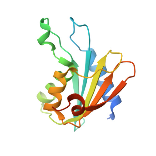 | |
UniProt | |||||
Find proteins for A0A6L7GZQ8 (Bacillus anthracis) Explore A0A6L7GZQ8 Go to UniProtKB: A0A6L7GZQ8 | |||||
Entity Groups | |||||
| Sequence Clusters | 30% Identity50% Identity70% Identity90% Identity95% Identity100% Identity | ||||
| UniProt Group | A0A6L7GZQ8 | ||||
Sequence AnnotationsExpand | |||||
| |||||
| Ligands 1 Unique | |||||
|---|---|---|---|---|---|
| ID | Chains | Name / Formula / InChI Key | 2D Diagram | 3D Interactions | |
| ZN Query on ZN | C [auth A], D [auth B] | ZINC ION Zn PTFCDOFLOPIGGS-UHFFFAOYSA-N |  | ||
| Length ( Å ) | Angle ( ˚ ) |
|---|---|
| a = 99.792 | α = 90 |
| b = 50.552 | β = 132.1 |
| c = 70.438 | γ = 90 |
| Software Name | Purpose |
|---|---|
| REFMAC | refinement |
| HKL-2000 | data reduction |
| SCALEPACK | data scaling |
| MOLREP | phasing |
RCSB PDB (citation) is hosted by
RCSB PDB is a member of the

