Crystal Structure of Atypical Cytoplasmic ABC-ATPase SufC from Thermus thermophilus HB8.
Watanabe, S., Kita, A., Miki, K.(2005) J Mol Biol 353: 1043-1054
- PubMed: 16216272
- DOI: https://doi.org/10.1016/j.jmb.2005.09.017
- Primary Citation of Related Structures:
2D2E, 2D2F - PubMed Abstract:
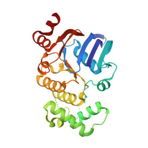
SufC, a cytoplasmic ABC-ATPase, is one of the most conserved Suf proteins. SufC forms a stable complex with SufB and SufD, and the SufBCD complex interacts with other Suf proteins in the Fe-S cluster assembly. We have determined the crystal structure of SufC from Thermus thermophilus HB8 in nucleotide-free and ADP-Mg-bound states at 1.7A and 1.9A resolution, respectively. The overall architecture of the SufC structure is similar to other ABC ATPases structures, but there are several specific motifs in SufC. Three residues following the end of the Walker B motif form a novel 3(10) helix which is not observed in other ABC ATPases. Due to the novel 3(10) helix, a conserved glutamate residue involved in ATP hydrolysis is flipped out. Although this unusual conformation is unfavorable for ATP hydrolysis, salt-bridges formed by conserved residues and a strong hydrogen-bonding network around the novel 3(10) helix suggest that the novel 3(10) helix of SufC is a rigid conserved motif. Compared to other ABC-ATPase structures, a significant displacement occurs at a linker region between the ABC alpha/beta domain and the alpha-helical domain. The linker conformation is stabilized by a hydrophobic interaction between conserved residues around the Q loop. The molecular surfaces of SufC and the C-terminal helices of SufD (PDB code: 1VH4) suggest that the unusual linker conformation conserved among SufC proteins is probably suitable for interacting with SufB and SufD.
Organizational Affiliation:
Department of Chemistry, Graduate School of Science, Kyoto University, Sakyo-ku, Kyoto 606-8502, Japan.