2CZV

Crystal structure of archeal RNase P protein ph1481p in complex with ph1877p


Experimental Data Snapshot

  • Method: X-RAY DIFFRACTION
  • Resolution: 2.00 Å
  • R-Value Free: 0.261 
  • R-Value Work: 0.218 
  • R-Value Observed: 0.218 

wwPDB Validation   3D Report Full Report


Ligand Structure Quality Assessment 


This is version 1.4 of the entry. See complete history


Literature

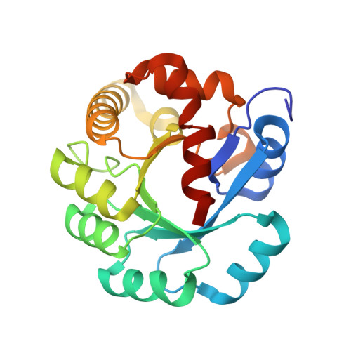
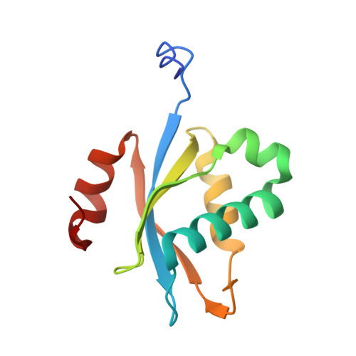
Crystal structure of protein Ph1481p in complex with protein Ph1877p of archaeal RNase P from Pyrococcus horikoshii OT3: implication of dimer formation of the holoenzyme

Kawano, S.Nakashima, T.Kakuta, Y.Tanaka, I.Kimura, M.

(2006) J Mol Biol 357: 583-591

  • DOI: https://doi.org/10.1016/j.jmb.2005.12.086
  • Primary Citation of Related Structures:  
    2CZV

  • PubMed Abstract: 

    Ribonuclease P (RNase P) in the hyperthermophilic archaeon Pyrococcus horikoshii OT3 consists of a catalytic RNA and five protein subunits. We previously determined crystal structures of four protein subunits. Ph1481p, an archaeal homologue for human hPop5, is the protein component of the P.horikoshii RNase P for which no structural information is available. Here we report the crystal structure of Ph1481p in complex with another protein subunit, Ph1877p, determined at 2.0 A resolution. Ph1481p consists of a five-stranded antiparallel beta-sheet and five helices, which fold in a way that is topologically similar to the ribonucleoprotein (RNP) domain. Ph1481p is, however, distinct from the typical RNP domain in that it has additional helices at the C terminus, which pack against one face of the beta-sheet. The presence of two complexes in the asymmetric unit, together with gel filtration chromatography indicates that the heterotetramer is stable in solution and represents a fundamental building block in the crystals. In the heterotetrameric structure (Ph1877p-(Ph1481p)(2)-Ph1877p), a homodimer of Ph1481p sits between two Ph1877p monomers. Ph1481p dimerizes through hydrogen bonding interaction from the loop between alpha1 and alpha2 helices, and each Ph1481p interacts with two Ph1877p molecules, where alpha2 and alpha3 in Ph1481p interact with alpha7 in one Ph1877p and alpha8 in the other Ph1877p molecule, respectively. Deletion of the alpha1-alpha2 loop in Ph1481p caused heterodimerization with Ph1877p, and abolished ability to homodimerize itself and heterotetramerize with Ph1877p. Furthermore, the reconstituted particle containing the deletion mutant Ph1481p (mPh1481p) exhibited significantly reduced nuclease activity. These results suggest the presence of the heterotetramer of Ph1481p and Ph1877p in P.horikoshii RNase P.


  • Organizational Affiliation

    Laboratory of Biochemistry, Department of Bioscience and Biotechnology, Faculty of Agriculture, Graduate School, Kyushu University, Fukuoka 812-8581, Japan.


Macromolecules
Find similar proteins by:  (by identity cutoff)  |  3D Structure
Entity ID: 1
MoleculeChains Sequence LengthOrganismDetailsImage
Ribonuclease P protein component 3
A, B
212Pyrococcus horikoshiiMutation(s): 0 
Gene Names: PH1877
EC: 3.1.26.5
UniProt
Find proteins for O59543 (Pyrococcus horikoshii (strain ATCC 700860 / DSM 12428 / JCM 9974 / NBRC 100139 / OT-3))
Explore O59543 
Go to UniProtKB:  O59543
Entity Groups  
Sequence Clusters30% Identity50% Identity70% Identity90% Identity95% Identity100% Identity
UniProt GroupO59543
Sequence Annotations
Expand
  • Reference Sequence
Find similar proteins by:  (by identity cutoff)  |  3D Structure
Entity ID: 2
MoleculeChains Sequence LengthOrganismDetailsImage
Ribonuclease P protein component 2
C, D
120Pyrococcus horikoshiiMutation(s): 1 
Gene Names: PH1481
EC: 3.1.26.5
UniProt
Find proteins for O59150 (Pyrococcus horikoshii (strain ATCC 700860 / DSM 12428 / JCM 9974 / NBRC 100139 / OT-3))
Explore O59150 
Go to UniProtKB:  O59150
Entity Groups  
Sequence Clusters30% Identity50% Identity70% Identity90% Identity95% Identity100% Identity
UniProt GroupO59150
Sequence Annotations
Expand
  • Reference Sequence
Small Molecules
Ligands 2 Unique
IDChains Name / Formula / InChI Key2D Diagram3D Interactions
BOG
Query on BOG

Download Ideal Coordinates CCD File 
E [auth A],
V [auth D]
octyl beta-D-glucopyranoside
C14 H28 O6
HEGSGKPQLMEBJL-RKQHYHRCSA-N
ACY
Query on ACY

Download Ideal Coordinates CCD File 
F [auth A]
G [auth A]
H [auth A]
I [auth A]
J [auth B]
F [auth A],
G [auth A],
H [auth A],
I [auth A],
J [auth B],
K [auth B],
L [auth B],
M [auth B],
N [auth C],
O [auth C],
P [auth C],
Q [auth C],
R [auth C],
S [auth C],
T [auth C],
U [auth C]
ACETIC ACID
C2 H4 O2
QTBSBXVTEAMEQO-UHFFFAOYSA-N
Experimental Data & Validation

Experimental Data

  • Method: X-RAY DIFFRACTION
  • Resolution: 2.00 Å
  • R-Value Free: 0.261 
  • R-Value Work: 0.218 
  • R-Value Observed: 0.218 
  • Space Group: P 21 21 21
Unit Cell:
Length ( Å )Angle ( ˚ )
a = 63.332α = 90
b = 76.118β = 90
c = 151.276γ = 90
Software Package:
Software NamePurpose
CNSrefinement
ADSCdata collection
SCALEPACKdata scaling
SOLVEphasing

Structure Validation

View Full Validation Report


Ligand Structure Quality Assessment 


Entry History 

Deposition Data

Revision History  (Full details and data files)

  • Version 1.0: 2006-06-27
    Type: Initial release
  • Version 1.1: 2008-04-30
    Changes: Version format compliance
  • Version 1.2: 2011-07-13
    Changes: Version format compliance
  • Version 1.3: 2020-07-29
    Type: Remediation
    Reason: Carbohydrate remediation
    Changes: Data collection, Derived calculations, Structure summary
  • Version 1.4: 2021-11-10
    Changes: Database references, Structure summary