2CTB

THE HIGH RESOLUTION CRYSTAL STRUCTURE OF THE COMPLEX BETWEEN CARBOXYPEPTIDASE A AND L-PHENYL LACTATE


Experimental Data Snapshot

  • Method: X-RAY DIFFRACTION
  • Resolution: 1.50 Å
  • R-Value Observed: 0.100 

wwPDB Validation   3D Report Full Report


This is version 1.3 of the entry. See complete history


Literature

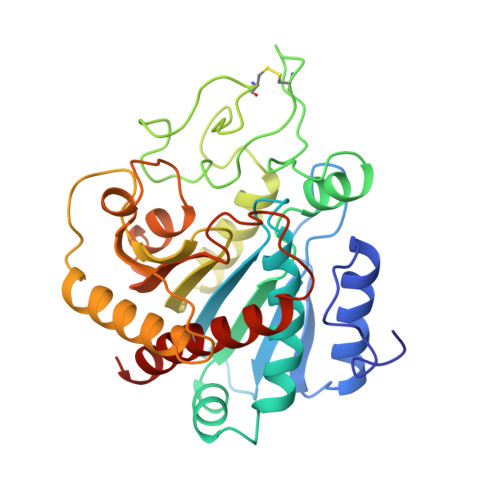
High-resolution structure of the complex between carboxypeptidase A and L-phenyl lactate.

Teplyakov, A.Wilson, K.S.Orioli, P.Mangani, S.

(1993) Acta Crystallogr D Biol Crystallogr 49: 534-540

  • DOI: https://doi.org/10.1107/S0907444993007267
  • Primary Citation of Related Structures:  
    2CTB, 2CTC

  • PubMed Abstract: 

    The X-ray structures of native carboxypeptidase A and of the enzyme-inhibitor complex with L-phenyl lactate have been refined at 1.54 and 1.45 A resolution to R factors of 0.151 and 0.161, respectively. Crystals of the complex were isomorphous with the native crystals (space group P2(1), a = 51.60, b = 60.27, c = 47.25 A, beta = 97.27 degrees ). The high-resolution electron density allowed correction of many side-chain positions in the classical carboxypeptidase A model. This reflects the advantages of the high-quality complete synchrotron data collected with an imaging plate detector. The conformational changes in the active centre of the enzyme upon binding of the inhibitor are restricted to only two residues, Tyr248 and Arg145. L-Phenyl lactate is bound in the S1' pocket and forms hydrogen bonds to Arg145, Glu270 and to the zinc-bound water molecule. The present structure provides an explanation for the higher stability of the complexes with the products of esterolysis in comparison with those of amidolysis. This is consistent with the finding that product release is rate limiting for esters but not for peptides.


  • Organizational Affiliation

    EMBL, c/o DESY, Hamburg, Germany.


Macromolecules
Find similar proteins by:  (by identity cutoff)  |  3D Structure
Entity ID: 1
MoleculeChains Sequence LengthOrganismDetailsImage
CARBOXYPEPTIDASE A307Bos taurusMutation(s): 0 
EC: 3.4.17.1
UniProt
Find proteins for P00730 (Bos taurus)
Explore P00730 
Go to UniProtKB:  P00730
Entity Groups  
Sequence Clusters30% Identity50% Identity70% Identity90% Identity95% Identity100% Identity
UniProt GroupP00730
Sequence Annotations
Expand
  • Reference Sequence
Small Molecules
Ligands 1 Unique
IDChains Name / Formula / InChI Key2D Diagram3D Interactions
ZN
Query on ZN

Download Ideal Coordinates CCD File 
B [auth A]ZINC ION
Zn
PTFCDOFLOPIGGS-UHFFFAOYSA-N
Experimental Data & Validation

Experimental Data

  • Method: X-RAY DIFFRACTION
  • Resolution: 1.50 Å
  • R-Value Observed: 0.100 
  • Space Group: P 1 21 1
Unit Cell:
Length ( Å )Angle ( ˚ )
a = 51.6α = 90
b = 60.27β = 97.27
c = 47.25γ = 90
Software Package:
Software NamePurpose
PROLSQrefinement

Structure Validation

View Full Validation Report


Entry History 

Deposition Data

Revision History  (Full details and data files)

  • Version 1.0: 1994-01-31
    Type: Initial release
  • Version 1.1: 2008-03-24
    Changes: Version format compliance
  • Version 1.2: 2011-07-13
    Changes: Version format compliance
  • Version 1.3: 2017-11-29
    Changes: Derived calculations, Other