Analysis of side-chain organization on a refined model of charybdotoxin: structural and functional implications.
Bontems, F., Gilquin, B., Roumestand, C., Menez, A., Toma, F.(1992) Biochemistry 31: 7756-7764
- PubMed: 1380828
- DOI: https://doi.org/10.1021/bi00149a003
- Primary Citation of Related Structures:
2CRD - PubMed Abstract:
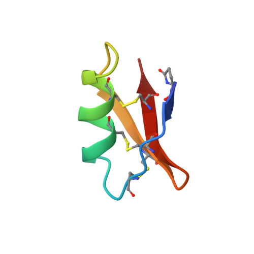
The spatial organization of side chains on a refined model of charybdotoxin is presented. First, the structural role of two groups of well-defined, low-accessible side chains (Thr3, Val5, Val16, Leu20, Cys33 and Leu20, His21, Thr23, Cys17, Cys35) is discussed. These side chains are conserved in three out of the five known scorpion toxins acting on K+ channels. Interestingly, they are not conserved in scyllatoxin which presents a slightly different secondary structure organization. Second, the spatial organization of all positively charged residues is analyzed. Comparison with the results presented by Park and Miller [(1992) Biochemistry (preceding paper in this issue)] shows that all functionally important positive residues are located on the beta-sheet side of the toxin. These results are different from those obtained by Auguste et al. [(1992) Biochemistry 31, 648-654] on scyllatoxin, which blocks a different type of K+ channel. This study shows, in fact, that functionally important positive residues are located on the helix side of the toxin. Thus, charybdotoxin and scyllatoxin, which present the same global fold, interact with two different classes of K+ channels by two different parts of the motif.
Organizational Affiliation:
Département d'Ingénierie et d'Etude des Protéines, CE-Saclay, Gif-sur-Yvette, France.