2CLB

The structure of the DPS-like protein from Sulfolobus solfataricus reveals a bacterioferritin-like di-metal binding site within a Dps- like dodecameric assembly


Experimental Data Snapshot

  • Method: X-RAY DIFFRACTION
  • Resolution: 2.40 Å
  • R-Value Free: 0.222 
  • R-Value Work: 0.199 
  • R-Value Observed: 0.200 

wwPDB Validation   3D Report Full Report


This is version 1.1 of the entry. See complete history


Literature

Structure of the Dps-Like Protein from Sulfolobus Solfataricus Reveals a Bacterioferritin-Like Dimetal Binding Site within a Dps-Like Dodecameric Assembly.

Gauss, G.H.Benas, P.Wiedenheft, B.Young, M.Douglas, T.Lawrence, C.M.

(2006) Biochemistry 45: 10815

  • DOI: https://doi.org/10.1021/bi060782u
  • Primary Citation of Related Structures:  
    2CLB

  • PubMed Abstract: 

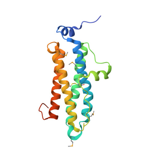
    The superfamily of ferritin-like proteins has recently expanded to include a phylogenetically distinct class of proteins termed DPS-like (DPSL) proteins. Despite their distinct genetic signatures, members of this subclass share considerable similarity to previously recognized DPS proteins. Like DPS, these proteins are expressed in response to oxidative stress, form dodecameric cage-like particles, preferentially utilize H(2)O(2) in the controlled oxidation of Fe(2+), and possess a short N-terminal extension implicated in stabilizing cellular DNA. Given these extensive similarities, the functional properties responsible for the preservation of the DPSL signature in the genomes of diverse prokaryotes have been unclear. Here, we describe the crystal structure of a DPSL protein from the thermoacidophilic archaeon Sulfolobus solfataricus. Although the overall fold of the polypeptide chain and the oligomeric state of this protein are indistinguishable from those of authentic DPS proteins, several important differences are observed. First, rather than a ferroxidase site at the subunit interface, as is observed in all other DPS proteins, the ferroxidase site in SsDPSL is buried within the four-helix bundle, similar to bacterioferritin. Second, the structure reveals a channel leading from the exterior surface of SsDPSL to the bacterioferritin-like dimetal binding site, possibly allowing divalent cations and/or H(2)O(2) to access the active site. Third, a pair of cysteine residues unique to DPSL proteins is found adjacent to the dimetal binding site juxtaposed between the exterior surface of the protein and the active site channel. The cysteine residues in this thioferritin motif may play a redox active role, possibly serving to recycle iron at the ferroxidase center.


  • Organizational Affiliation

    Thermal Biology Institute, Montana State University, Bozeman, Montana 59717, USA.


Macromolecules
Find similar proteins by:  (by identity cutoff)  |  3D Structure
Entity ID: 1
MoleculeChains Sequence LengthOrganismDetailsImage
DPS-LIKE PROTEIN188Saccharolobus solfataricus P2Mutation(s): 0 
UniProt
Find proteins for P95855 (Saccharolobus solfataricus (strain ATCC 35092 / DSM 1617 / JCM 11322 / P2))
Explore P95855 
Go to UniProtKB:  P95855
Entity Groups  
Sequence Clusters30% Identity50% Identity70% Identity90% Identity95% Identity100% Identity
UniProt GroupP95855
Sequence Annotations
Expand
  • Reference Sequence
Small Molecules
Ligands 2 Unique
IDChains Name / Formula / InChI Key2D Diagram3D Interactions
ZN
Query on ZN

Download Ideal Coordinates CCD File 
I [auth A]
K [auth B]
M [auth C]
O [auth D]
Q [auth M]
I [auth A],
K [auth B],
M [auth C],
O [auth D],
Q [auth M],
S [auth N],
U [auth O],
W [auth P]
ZINC ION
Zn
PTFCDOFLOPIGGS-UHFFFAOYSA-N
FE
Query on FE

Download Ideal Coordinates CCD File 
J [auth A]
L [auth B]
N [auth C]
P [auth D]
R [auth M]
J [auth A],
L [auth B],
N [auth C],
P [auth D],
R [auth M],
T [auth N],
V [auth O],
X [auth P]
FE (III) ION
Fe
VTLYFUHAOXGGBS-UHFFFAOYSA-N
Modified Residues  1 Unique
IDChains TypeFormula2D DiagramParent
MSE
Query on MSE
A
B
C
D
E [auth M]
A,
B,
C,
D,
E [auth M],
F [auth N],
G [auth O],
H [auth P]
L-PEPTIDE LINKINGC5 H11 N O2 SeMET
Experimental Data & Validation

Experimental Data

  • Method: X-RAY DIFFRACTION
  • Resolution: 2.40 Å
  • R-Value Free: 0.222 
  • R-Value Work: 0.199 
  • R-Value Observed: 0.200 
  • Space Group: I 21 3
Unit Cell:
Length ( Å )Angle ( ˚ )
a = 212.928α = 90
b = 212.928β = 90
c = 212.928γ = 90
Software Package:
Software NamePurpose
REFMACrefinement
HKL-2000data reduction
HKL-2000data scaling
SOLVEphasing

Structure Validation

View Full Validation Report


Entry History 

Deposition Data

Revision History  (Full details and data files)

  • Version 1.0: 2006-07-17
    Type: Initial release
  • Version 1.1: 2011-07-13
    Changes: Advisory, Version format compliance