2CJW

Crystal structure of the small GTPase Gem (GemDNDCaM) in complex to Mg.GDP


Experimental Data Snapshot

  • Method: X-RAY DIFFRACTION
  • Resolution: 2.10 Å
  • R-Value Free: 0.241 
  • R-Value Work: 0.209 
  • R-Value Observed: 0.212 

wwPDB Validation   3D Report Full Report


Ligand Structure Quality Assessment 


This is version 1.3 of the entry. See complete history


Literature

Biochemical and Structural Characterization of the Gem Gtpase.

Splingard, A.Menetrey, J.Perderiset, M.Cicolari, J.Regazzoni, K.Hamoudi, F.Cabanie, L.El Marjou, A.Wells, A.Houdusse, A.De Gunzburg, J.

(2007) J Biol Chem 282: 1905

  • DOI: https://doi.org/10.1074/jbc.M604363200
  • Primary Citation of Related Structures:  
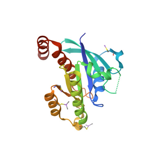
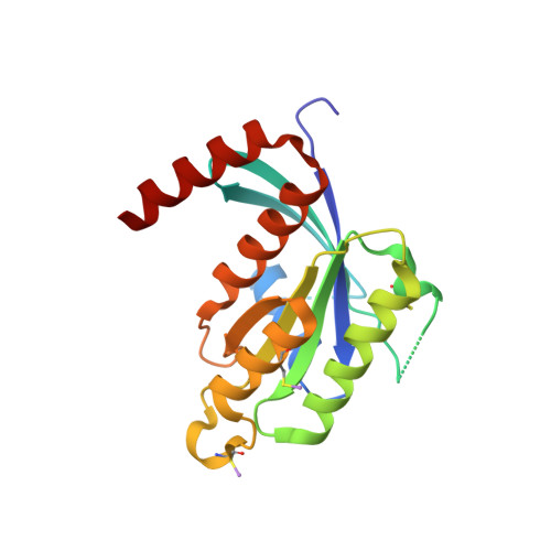
    2CJW

  • PubMed Abstract: 

    RGK proteins, encompassing Rad, Gem, Rem1, and Rem2, constitute an intriguing branch of the Ras superfamily; their expression is regulated at the transcription level, they exhibit atypical nucleotide binding motifs, and they carry both large N- and C-terminal extensions. Biochemical and structural studies are required to better understand how such proteins function. Here, we report the first structure for a RGK protein: the crystal structure of a truncated form of the human Gem protein (G domain plus the first part of the C-terminal extension) in complex with Mg.GDP at 2.1 A resolution. It reveals that the G-domain fold and Mg.GDP binding site of Gem are similar to those found for other Ras family GTPases. The first part of the C-terminal extension adopts an alpha-helical conformation that extends along the alpha5 helix and interacts with the tip of the interswitch. Biochemical studies show that the affinities of Gem for GDP and GTP are considerably lower (micromolar range) compared with H-Ras, independent of the presence or absence of N- and C-terminal extensions, whereas its GTPase activity is higher than that of H-Ras and regulated by both extensions. We show how the bulky DXWEX motif, characteristic of the switch II of RGK proteins, affects the conformation of switch I and the phosphate-binding site. Altogether, our data reveal that Gem is a bona fide GTPase that exhibits striking structural and biochemical features that should impact its regulation and cellular activities.


  • Organizational Affiliation

    Institut Curie, Centre de Recherche, Paris F-75248, France, INSERM U528, Paris F-75248, France.


Macromolecules
Find similar proteins by:  (by identity cutoff)  |  3D Structure
Entity ID: 1
MoleculeChains Sequence LengthOrganismDetailsImage
GTP-BINDING PROTEIN GEM192Homo sapiensMutation(s): 0 
UniProt & NIH Common Fund Data Resources
Find proteins for P55040 (Homo sapiens)
Explore P55040 
Go to UniProtKB:  P55040
PHAROS:  P55040
GTEx:  ENSG00000164949 
Entity Groups  
Sequence Clusters30% Identity50% Identity70% Identity90% Identity95% Identity100% Identity
UniProt GroupP55040
Sequence Annotations
Expand
  • Reference Sequence
Find similar proteins by:  (by identity cutoff)  |  3D Structure
Entity ID: 2
MoleculeChains Sequence LengthOrganismDetailsImage
GTP-BINDING PROTEIN GEM192Homo sapiensMutation(s): 0 
UniProt & NIH Common Fund Data Resources
Find proteins for P55040 (Homo sapiens)
Explore P55040 
Go to UniProtKB:  P55040
PHAROS:  P55040
GTEx:  ENSG00000164949 
Entity Groups  
Sequence Clusters30% Identity50% Identity70% Identity90% Identity95% Identity100% Identity
UniProt GroupP55040
Sequence Annotations
Expand
  • Reference Sequence
Small Molecules
Modified Residues  1 Unique
IDChains TypeFormula2D DiagramParent
CAS
Query on CAS
A
L-PEPTIDE LINKINGC5 H12 As N O2 SCYS
Binding Affinity Annotations 
IDSourceBinding Affinity
GDP Binding MOAD:  2CJW Kd: 2500 (nM) from 1 assay(s)
Experimental Data & Validation

Experimental Data

  • Method: X-RAY DIFFRACTION
  • Resolution: 2.10 Å
  • R-Value Free: 0.241 
  • R-Value Work: 0.209 
  • R-Value Observed: 0.212 
  • Space Group: P 61
Unit Cell:
Length ( Å )Angle ( ˚ )
a = 116.617α = 90
b = 116.617β = 90
c = 81.409γ = 120
Software Package:
Software NamePurpose
REFMACrefinement
MOSFLMdata reduction
SCALAdata scaling
AMoREphasing

Structure Validation

View Full Validation Report


Ligand Structure Quality Assessment 


Entry History 

Deposition Data

Revision History  (Full details and data files)

  • Version 1.0: 2006-11-09
    Type: Initial release
  • Version 1.1: 2011-05-08
    Changes: Version format compliance
  • Version 1.2: 2011-07-13
    Changes: Version format compliance
  • Version 1.3: 2023-12-13
    Changes: Data collection, Database references, Derived calculations, Other, Refinement description