2CI1

Crystal Structure of dimethylarginine dimethylaminohydrolase I in complex with S-nitroso-Lhomocysteine


Experimental Data Snapshot

  • Method: X-RAY DIFFRACTION
  • Resolution: 1.08 Å
  • R-Value Free: 0.141 

wwPDB Validation   3D Report Full Report


This is version 1.3 of the entry. See complete history


Literature

Structure of the Mammalian Nos Regulator Dimethylarginine Dimethylaminohydrolase: A Basis for the Design of Specific Inhibitors.

Frey, D.Braun, O.Briand, C.Vasak, M.Grutter, M.G.

(2006) Structure 14: 901

  • DOI: https://doi.org/10.1016/j.str.2006.03.006
  • Primary Citation of Related Structures:  
    2C6Z, 2CI1, 2CI3, 2CI4, 2CI5, 2CI6, 2CI7

  • PubMed Abstract: 

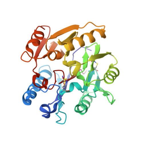
    Dimethylarginine dimethylaminohydrolase (DDAH) is involved in the regulation of nitric oxide synthase (NOS) by metabolizing the free endogenous arginine derivatives N(omega)-methyl-L-arginine (MMA) and N(omega),N(omega)-dimethyl-L-arginine (ADMA), which are competitive inhibitors of NOS. Here, we present high-resolution crystal structures of DDAH isoform 1 (DDAH-1) isolated from bovine brain in complex with different inhibitors, including S-nitroso-L-homocysteine and Zn2+, a regulator of this mammalian enzyme. The structure of DDAH-1 consists of a propeller-like fold similar to other arginine-modifying enzymes and a flexible loop, which adopts different conformations and acts as a lid at the entrance of the active site. The orientation and interaction mode of inhibitors in the active site give insight into the regulation and the molecular mechanism of the enzyme. The presented structures provide a basis for the structure-based development of specific DDAH-1 inhibitors that might be useful in the therapeutic treatment of NOS dysfunction-related diseases.


  • Organizational Affiliation

    Department of Biochemistry, University of Zürich, Winterthurerstrasse 190, CH-8057 Zürich, Switzerland.


Macromolecules
Find similar proteins by:  (by identity cutoff)  |  3D Structure
Entity ID: 1
MoleculeChains Sequence LengthOrganismDetailsImage
NG, NG-DIMETHYLARGININE DIMETHYLAMINOHYDROLASE 1275Bos taurusMutation(s): 2 
EC: 3.5.3.18
UniProt
Find proteins for P56965 (Bos taurus)
Explore P56965 
Go to UniProtKB:  P56965
Entity Groups  
Sequence Clusters30% Identity50% Identity70% Identity90% Identity95% Identity100% Identity
UniProt GroupP56965
Sequence Annotations
Expand
  • Reference Sequence
Small Molecules
Ligands 1 Unique
IDChains Name / Formula / InChI Key2D Diagram3D Interactions
CIT
Query on CIT

Download Ideal Coordinates CCD File 
B [auth A]CITRIC ACID
C6 H8 O7
KRKNYBCHXYNGOX-UHFFFAOYSA-N
Modified Residues  3 Unique
IDChains TypeFormula2D DiagramParent
K1R
Query on K1R
A
L-PEPTIDE LINKINGC7 H15 N3 O5 S2CYS
KOR
Query on KOR
A
L-PEPTIDE LINKINGC7 H15 N3 O5 S2MET
SNC
Query on SNC
A
L-PEPTIDE LINKINGC3 H6 N2 O3 SCYS
Experimental Data & Validation

Experimental Data

  • Method: X-RAY DIFFRACTION
  • Resolution: 1.08 Å
  • R-Value Free: 0.141 
  • Space Group: P 1 21 1
Unit Cell:
Length ( Å )Angle ( ˚ )
a = 40.664α = 90
b = 79.91β = 108.58
c = 44.894γ = 90
Software Package:
Software NamePurpose
SHELXL-97refinement
DENZOdata reduction
SCALEPACKdata scaling
AMoREphasing

Structure Validation

View Full Validation Report


Entry History 

Deposition Data

Revision History  (Full details and data files)

  • Version 1.0: 2006-05-17
    Type: Initial release
  • Version 1.1: 2011-08-10
    Changes: Derived calculations, Non-polymer description, Other, Refinement description, Version format compliance
  • Version 1.2: 2019-05-22
    Changes: Data collection, Derived calculations, Other, Refinement description
  • Version 1.3: 2023-12-13
    Changes: Data collection, Database references, Derived calculations, Other, Refinement description