2CDQ

Crystal structure of Arabidopsis thaliana aspartate kinase complexed with lysine and S- adenosylmethionine


Experimental Data Snapshot

  • Method: X-RAY DIFFRACTION
  • Resolution: 2.85 Å
  • R-Value Free: 0.244 
  • R-Value Work: 0.202 
  • R-Value Observed: 0.204 

wwPDB Validation   3D Report Full Report


Ligand Structure Quality Assessment 


This is version 1.1 of the entry. See complete history


Literature

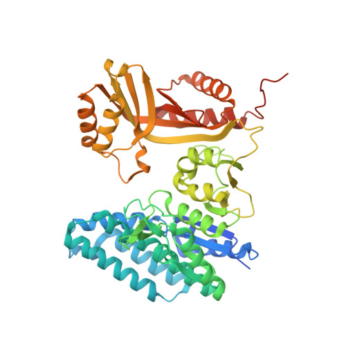
A Novel Organization of Act Domains in Allosteric Enzymes Revealed by the Crystal Structure of Arabidopsis Aspartate Kinase

Mas-Droux, C.Curien, G.Robert-Genthon, M.Laurencin, M.Ferrer, J.L.Dumas, R.

(2006) Plant Cell 18: 1681

  • DOI: https://doi.org/10.1105/tpc.105.040451
  • Primary Citation of Related Structures:  
    2CDQ

  • PubMed Abstract: 

    Asp kinase catalyzes the first step of the Asp-derived essential amino acid pathway in plants and microorganisms. Depending on the source organism, this enzyme contains up to four regulatory ACT domains and exhibits several isoforms under the control of a great variety of allosteric effectors. We report here the dimeric structure of a Lys and S-adenosylmethionine-sensitive Asp kinase isoform from Arabidopsis thaliana in complex with its two inhibitors. This work reveals the structure of an Asp kinase and an enzyme containing two ACT domains cocrystallized with its effectors. Only one ACT domain (ACT1) is implicated in effector binding. A loop involved in the binding of Lys and S-adenosylmethionine provides an explanation for the synergistic inhibition by these effectors. The presence of S-adenosylmethionine in the regulatory domain indicates that ACT domains are also able to bind nucleotides. The organization of ACT domains in the present structure is different from that observed in Thr deaminase and in the regulatory subunit of acetohydroxyacid synthase III.


  • Organizational Affiliation

    Centre National de la Recherche Scientifique, Institut National de la Recherche Agronomique, Université Joseph Fourier, Commissariat à l'Energie Atomique, Département Réponse et Dynamique Cellulaires, France.


Macromolecules
Find similar proteins by:  (by identity cutoff)  |  3D Structure
Entity ID: 1
MoleculeChains Sequence LengthOrganismDetailsImage
ASPARTOKINASE
A, B
510Arabidopsis thalianaMutation(s): 0 
EC: 2.7.2.4
UniProt
Find proteins for Q9LYU8 (Arabidopsis thaliana)
Explore Q9LYU8 
Go to UniProtKB:  Q9LYU8
Entity Groups  
Sequence Clusters30% Identity50% Identity70% Identity90% Identity95% Identity100% Identity
UniProt GroupQ9LYU8
Sequence Annotations
Expand
  • Reference Sequence
Small Molecules
Ligands 3 Unique
IDChains Name / Formula / InChI Key2D Diagram3D Interactions
SAM
Query on SAM

Download Ideal Coordinates CCD File 
F [auth A],
J [auth B]
S-ADENOSYLMETHIONINE
C15 H22 N6 O5 S
MEFKEPWMEQBLKI-FCKMPRQPSA-N
TAR
Query on TAR

Download Ideal Coordinates CCD File 
E [auth A],
I [auth B]
D(-)-TARTARIC ACID
C4 H6 O6
FEWJPZIEWOKRBE-LWMBPPNESA-N
LYS
Query on LYS

Download Ideal Coordinates CCD File 
C [auth A],
D [auth A],
G [auth B],
H [auth B]
LYSINE
C6 H15 N2 O2
KDXKERNSBIXSRK-YFKPBYRVSA-O
Experimental Data & Validation

Experimental Data

  • Method: X-RAY DIFFRACTION
  • Resolution: 2.85 Å
  • R-Value Free: 0.244 
  • R-Value Work: 0.202 
  • R-Value Observed: 0.204 
  • Space Group: I 41
Unit Cell:
Length ( Å )Angle ( ˚ )
a = 117.342α = 90
b = 117.342β = 90
c = 255.283γ = 90
Software Package:
Software NamePurpose
REFMACrefinement
MOSFLMdata reduction
SCALAdata scaling
MOLREPphasing

Structure Validation

View Full Validation Report


Ligand Structure Quality Assessment 


Entry History 

Deposition Data

Revision History  (Full details and data files)

  • Version 1.0: 2006-05-30
    Type: Initial release
  • Version 1.1: 2011-07-13
    Changes: Advisory, Version format compliance