2CDN

Crystal structure of Mycobacterium tuberculosis adenylate kinase complexed with two molecules of ADP and Mg


Experimental Data Snapshot

  • Method: X-RAY DIFFRACTION
  • Resolution: 1.90 Å
  • R-Value Free: 0.214 
  • R-Value Work: 0.169 
  • R-Value Observed: 0.172 

wwPDB Validation   3D Report Full Report


Ligand Structure Quality Assessment 


This is version 1.3 of the entry. See complete history


Literature

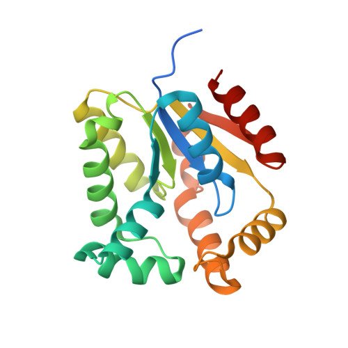
The Crystal Structure of Mycobacterium Tuberculosis Adenylate Kinase in Complex with Two Molecules of Adp and Mg2+ Supports an Associative Mechanism for Phosphoryl Transfer.

Bellinzoni, M.Haouz, A.Grana, M.Munier-Lehmann, H.Shepard, W.Alzari, P.M.

(2006) Protein Sci 15: 1489

  • DOI: https://doi.org/10.1110/ps.062163406
  • Primary Citation of Related Structures:  
    2CDN

  • PubMed Abstract: 

    The crystal structure of Mycobacterium tuberculosis adenylate kinase (MtAK) in complex with two ADP molecules and Mg2+ has been determined at 1.9 A resolution. Comparison with the solution structure of the enzyme, obtained in the absence of substrates, shows significant conformational changes of the LID and NMP-binding domains upon substrate binding. The ternary complex represents the state of the enzyme at the start of the backward reaction (ATP synthesis). The structure is consistent with a direct nucleophilic attack of a terminal oxygen from the acceptor ADP molecule on the beta-phosphate from the donor substrate, and both the geometry and the distribution of positive charge in the active site support the hypothesis of an associative mechanism for phosphoryl transfer.


  • Organizational Affiliation

    Unité de Biochimie Structurale, CNRS-URA 2185, Institut Pasteur, F-75724 Paris, France.


Macromolecules
Find similar proteins by:  (by identity cutoff)  |  3D Structure
Entity ID: 1
MoleculeChains Sequence LengthOrganismDetailsImage
ADENYLATE KINASE201Mycobacterium tuberculosis H37RvMutation(s): 0 
EC: 2.7.4.3
UniProt
Find proteins for P9WKF5 (Mycobacterium tuberculosis (strain ATCC 25618 / H37Rv))
Explore P9WKF5 
Go to UniProtKB:  P9WKF5
Entity Groups  
Sequence Clusters30% Identity50% Identity70% Identity90% Identity95% Identity100% Identity
UniProt GroupP9WKF5
Sequence Annotations
Expand
  • Reference Sequence
Experimental Data & Validation

Experimental Data

  • Method: X-RAY DIFFRACTION
  • Resolution: 1.90 Å
  • R-Value Free: 0.214 
  • R-Value Work: 0.169 
  • R-Value Observed: 0.172 
  • Space Group: P 1 21 1
Unit Cell:
Length ( Å )Angle ( ˚ )
a = 36.544α = 90
b = 64.91β = 110.12
c = 39.773γ = 90
Software Package:
Software NamePurpose
REFMACrefinement
MOSFLMdata reduction
SCALAdata scaling
AMoREphasing

Structure Validation

View Full Validation Report


Ligand Structure Quality Assessment 


Entry History 

Deposition Data

Revision History  (Full details and data files)

  • Version 1.0: 2006-05-10
    Type: Initial release
  • Version 1.1: 2011-05-08
    Changes: Version format compliance
  • Version 1.2: 2011-07-13
    Changes: Version format compliance
  • Version 1.3: 2023-12-13
    Changes: Data collection, Database references, Derived calculations, Other, Refinement description