2CBZ

Structure of the human Multidrug Resistance Protein 1 Nucleotide Binding Domain 1


Experimental Data Snapshot

  • Method: X-RAY DIFFRACTION
  • Resolution: 1.50 Å
  • R-Value Free: 0.187 
  • R-Value Work: 0.160 
  • R-Value Observed: 0.161 

wwPDB Validation   3D Report Full Report


Ligand Structure Quality Assessment 


This is version 1.3 of the entry. See complete history


Literature

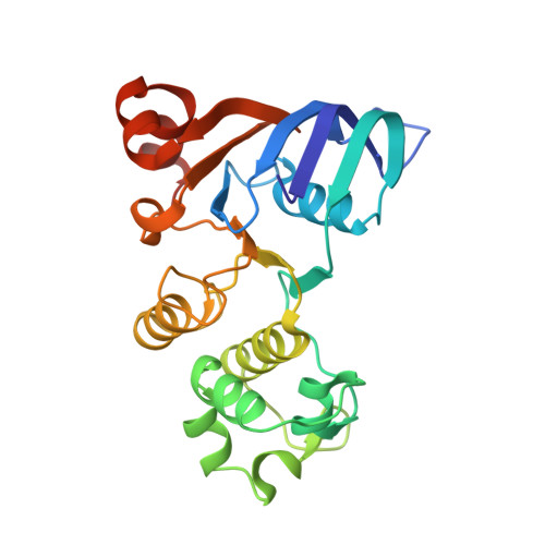
Structure of the Human Multidrug Resistance Protein 1 Nucleotide Binding Domain 1 Bound to Mg(2+)/ATP Reveals a Non-Productive Catalytic Site.

Ramaen, O.Leulliot, N.Sizun, C.Ulryck, N.Pamlard, O.Lallemand, J.-Y.Van Tilbeurgh, H.Jacquet, E.

(2006) J Mol Biol 359: 940

  • DOI: https://doi.org/10.1016/j.jmb.2006.04.005
  • Primary Citation of Related Structures:  
    2CBZ

  • PubMed Abstract: 

    Human multidrug resistance protein 1 (MRP1) is a membrane protein that belongs to the ATP-binding cassette (ABC) superfamily of transport proteins. MRP1 contributes to chemotherapy failure by exporting a wide range of anti-cancer drugs when over expressed in the plasma membrane of cells. Here, we report the first high-resolution crystal structure of human MRP1-NBD1. Drug efflux requires energy resulting from hydrolysis of ATP by nucleotide binding domains (NBDs). Contrary to the prokaryotic NBDs, the extremely low intrinsic ATPase activity of isolated MRP1-NBDs allowed us to obtain the structure of wild-type NBD1 in complex with Mg2+/ATP. The structure shows that MRP1-NBD1 adopts a canonical fold, but reveals an unexpected non-productive conformation of the catalytic site, providing an explanation for the low intrinsic ATPase activity of NBD1 and new hypotheses on the cooperativity of ATPase activity between NBD1 and NBD2 upon heterodimer formation.


  • Organizational Affiliation

    Institut de Chimie des Substances Naturelles, UPR 2301, Centre National de la Recherche Scientifique, Avenue de la Terrasse, 91190 Gif-sur-Yvette, France.


Macromolecules
Find similar proteins by:  (by identity cutoff)  |  3D Structure
Entity ID: 1
MoleculeChains Sequence LengthOrganismDetailsImage
MULTIDRUG RESISTANCE-ASSOCIATED PROTEIN 1237Homo sapiensMutation(s): 0 
UniProt & NIH Common Fund Data Resources
Find proteins for P33527 (Homo sapiens)
Explore P33527 
Go to UniProtKB:  P33527
PHAROS:  P33527
GTEx:  ENSG00000103222 
Entity Groups  
Sequence Clusters30% Identity50% Identity70% Identity90% Identity95% Identity100% Identity
UniProt GroupP33527
Sequence Annotations
Expand
  • Reference Sequence
Small Molecules
Ligands 2 Unique
IDChains Name / Formula / InChI Key2D Diagram3D Interactions
ATP
Query on ATP

Download Ideal Coordinates CCD File 
C [auth A]ADENOSINE-5'-TRIPHOSPHATE
C10 H16 N5 O13 P3
ZKHQWZAMYRWXGA-KQYNXXCUSA-N
MG
Query on MG

Download Ideal Coordinates CCD File 
B [auth A]MAGNESIUM ION
Mg
JLVVSXFLKOJNIY-UHFFFAOYSA-N
Binding Affinity Annotations 
IDSourceBinding Affinity
ATP PDBBind:  2CBZ Kd: 1.18e+5 (nM) from 1 assay(s)
Binding MOAD:  2CBZ Kd: 1.18e+5 (nM) from 1 assay(s)
Experimental Data & Validation

Experimental Data

  • Method: X-RAY DIFFRACTION
  • Resolution: 1.50 Å
  • R-Value Free: 0.187 
  • R-Value Work: 0.160 
  • R-Value Observed: 0.161 
  • Space Group: P 31
Unit Cell:
Length ( Å )Angle ( ˚ )
a = 65.56α = 90
b = 65.56β = 90
c = 64.612γ = 120
Software Package:
Software NamePurpose
REFMACrefinement
MOSFLMdata reduction
SCALAdata scaling
MOLREPphasing

Structure Validation

View Full Validation Report


Ligand Structure Quality Assessment 


Entry History 

Deposition Data

Revision History  (Full details and data files)

  • Version 1.0: 2006-05-17
    Type: Initial release
  • Version 1.1: 2011-05-08
    Changes: Version format compliance
  • Version 1.2: 2011-07-13
    Changes: Version format compliance
  • Version 1.3: 2023-12-13
    Changes: Data collection, Database references, Derived calculations, Other, Refinement description