2CBR

CELLULAR RETINOIC ACID BINDING PROTEIN I IN COMPLEX WITH A RETINOBENZOIC ACID (AM80)


Experimental Data Snapshot

  • Method: X-RAY DIFFRACTION
  • Resolution: 2.80 Å
  • R-Value Free: 0.268 
  • R-Value Work: 0.230 

wwPDB Validation   3D Report Full Report


Ligand Structure Quality Assessment 


This is version 1.5 of the entry. See complete history


Literature

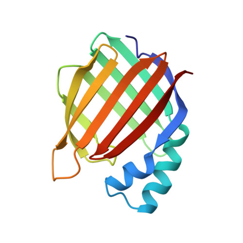
Structures of cellular retinoic acid binding proteins I and II in complex with synthetic retinoids.

Chaudhuri, B.N.Kleywegt, G.J.Broutin-L'Hermite, I.Bergfors, T.Senn, H.Le Motte, P.Partouche, O.Jones, T.A.

(1999) Acta Crystallogr D Biol Crystallogr 55: 1850-1857

  • DOI: https://doi.org/10.1107/s0907444999011026
  • Primary Citation of Related Structures:  
    2CBR, 2CBS, 3CBS

  • PubMed Abstract: 

    Retinoids play important roles in diverse cellular processes including growth, cell differentiation and vision. Many natural and synthetic retinoids are used as drugs in dermatology and oncology. A large amount of data has been accumulated on the cellular activity of different synthetic retinoids. They are stabilized and transported inside the cell cytoplasm by binding and transport proteins, such as cellular retinol-binding proteins and cellular retinoic acid binding proteins (CRABPs). The structures of human CRABP II in complex with two different synthetic retinoids, Ro13-6307 and Ro12--7310 (at 2.1 and 2.0 A resolution, respectively) and of bovine CRABP I in complex with a retinobenzoic acid, Am80 (at 2.8 A resolution) are described. The binding affinities of human CRABP I and II for the retinoids studied here have been determined. All these compounds have comparable binding affinities (nanomolar range) for both CRABPs. Apart from the particular interactions of the carboxylate group of the retinoids with specific protein groups, each structure reveals characteristic interactions. Studying the atomic details of the interaction of retinoids with retinoid-binding proteins facilitates the understanding of the kinetics of retinoid trafficking inside the cytoplasm.


  • Organizational Affiliation

    Department of Cell Biology, Uppsala University, Biomedical Center, Box 596, SE-751 24, Uppsala, Sweden.


Macromolecules
Find similar proteins by:  (by identity cutoff)  |  3D Structure
Entity ID: 1
MoleculeChains Sequence LengthOrganismDetailsImage
PROTEIN (CRABP-I)136Bos taurusMutation(s): 0 
Gene Names: MOUSE
UniProt
Find proteins for P62964 (Bos taurus)
Explore P62964 
Go to UniProtKB:  P62964
Entity Groups  
Sequence Clusters30% Identity50% Identity70% Identity90% Identity95% Identity100% Identity
UniProt GroupP62964
Sequence Annotations
Expand
  • Reference Sequence
Small Molecules
Ligands 1 Unique
IDChains Name / Formula / InChI Key2D Diagram3D Interactions
A80
Query on A80

Download Ideal Coordinates CCD File 
B [auth A]4-[(5,5,8,8-tetramethyl-5,6,7,8-tetrahydronaphthalen-2-yl)carbamoyl]benzoic acid
C22 H25 N O3
MUTNCGKQJGXKEM-UHFFFAOYSA-N
Binding Affinity Annotations 
IDSourceBinding Affinity
A80 PDBBind:  2CBR IC50: 140 (nM) from 1 assay(s)
Experimental Data & Validation

Experimental Data

  • Method: X-RAY DIFFRACTION
  • Resolution: 2.80 Å
  • R-Value Free: 0.268 
  • R-Value Work: 0.230 
  • Space Group: P 32 2 1
Unit Cell:
Length ( Å )Angle ( ˚ )
a = 133.5α = 90
b = 133.5β = 90
c = 40.57γ = 120
Software Package:
Software NamePurpose
DENZOdata reduction
SCALAdata scaling
AMoREphasing
CNSrefinement
CCP4data scaling

Structure Validation

View Full Validation Report


Ligand Structure Quality Assessment 


Entry History 

Deposition Data

Revision History  (Full details and data files)

  • Version 1.0: 1999-12-21
    Type: Initial release
  • Version 1.1: 2008-04-26
    Changes: Version format compliance
  • Version 1.2: 2011-07-13
    Changes: Version format compliance
  • Version 1.3: 2011-10-05
    Changes: Non-polymer description
  • Version 1.4: 2019-11-06
    Changes: Data collection, Database references
  • Version 1.5: 2023-08-23
    Changes: Data collection, Database references, Derived calculations, Refinement description