2C9B

Lumazine Synthase from Mycobacterium tuberculosus Bound to 3-(1,3,7- TRIHYDRO-9-D-RIBITYL-2,6,8-PURINETRIONE-7-YL)


Experimental Data Snapshot

  • Method: X-RAY DIFFRACTION
  • Resolution: 2.75 Å
  • R-Value Free: 0.324 
  • R-Value Work: 0.250 
  • R-Value Observed: 0.254 

wwPDB Validation   3D Report Full Report


Ligand Structure Quality Assessment 


This is version 1.3 of the entry. See complete history


Literature

Structural and Thermodynamic Insights Into the Binding Mode of Five Novel Inhibitors of Lumazine Synthase from Mycobacterium Tuberculosis.

Morgunova, E.Illarionov, B.Sambaiah, T.Haase, I.Bacher, A.Cushman, M.Fischer, M.Ladenstein, R.

(2006) FEBS J 273: 4790-4804

  • DOI: https://doi.org/10.1111/j.1742-4658.2006.05481.x
  • Primary Citation of Related Structures:  
    2C92, 2C94, 2C97, 2C9B, 2C9D

  • PubMed Abstract: 

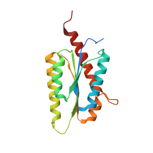
    Recently published genomic investigations of the human pathogen Mycobacterium tuberculosis have revealed that genes coding the proteins involved in riboflavin biosynthesis are essential for the growth of the organism. Because the enzymes involved in cofactor biosynthesis pathways are not present in humans, they appear to be promising candidates for the development of therapeutic drugs. The substituted purinetrione compounds have demonstrated high affinity and specificity to lumazine synthase, which catalyzes the penultimate step of riboflavin biosynthesis in bacteria and plants. The structure of M. tuberculosis lumazine synthase in complex with five different inhibitor compounds is presented, together with studies of the binding reactions by isothermal titration calorimetry. The inhibitors showed the association constants in the micromolar range. The analysis of the structures demonstrated the specific features of the binding of different inhibitors. The comparison of the structures and binding modes of five different inhibitors allows us to propose the ribitylpurinetrione compounds with C4-C5 alkylphosphate chains as most promising leads for further development of therapeutic drugs against M. tuberculosis.


  • Organizational Affiliation

    Karolinska Institutet, NOVUM, Centre for Structural Biochemistry, Huddinge, Sweden. katja.morgunova@biosci.ki.se


Macromolecules
Find similar proteins by:  (by identity cutoff)  |  3D Structure
Entity ID: 1
MoleculeChains Sequence LengthOrganismDetailsImage
6,7-DIMETHYL-8-RIBITYLLUMAZINE SYNTHASE
A, B, C, D, E
A, B, C, D, E, F, G, H, I, J
160Mycobacterium tuberculosisMutation(s): 0 
EC: 2.5.1.9
UniProt
Find proteins for P9WHE9 (Mycobacterium tuberculosis (strain ATCC 25618 / H37Rv))
Explore P9WHE9 
Go to UniProtKB:  P9WHE9
Entity Groups  
Sequence Clusters30% Identity50% Identity70% Identity90% Identity95% Identity100% Identity
UniProt GroupP9WHE9
Sequence Annotations
Expand
  • Reference Sequence
Small Molecules
Ligands 4 Unique
IDChains Name / Formula / InChI Key2D Diagram3D Interactions
PUG
Query on PUG

Download Ideal Coordinates CCD File 
AA [auth E]
EA [auth F]
HA [auth G]
K [auth A]
LA [auth H]
AA [auth E],
EA [auth F],
HA [auth G],
K [auth A],
LA [auth H],
O [auth B],
OA [auth I],
R [auth C],
RA [auth J],
W [auth D]
3-(1,3,7-TRIHYDRO-9-D-RIBITYL-2,6,8-PURINETRIONE-7-YL)
C10 H14 N4 O7
XIBHHWBJHOTHGZ-KODRXGBYSA-N
MPD
Query on MPD

Download Ideal Coordinates CCD File 
UA [auth J],
V [auth C]
(4S)-2-METHYL-2,4-PENTANEDIOL
C6 H14 O2
SVTBMSDMJJWYQN-YFKPBYRVSA-N
PO4
Query on PO4

Download Ideal Coordinates CCD File 
DA [auth E]
GA [auth F]
KA [auth G]
N [auth A]
NA [auth H]
DA [auth E],
GA [auth F],
KA [auth G],
N [auth A],
NA [auth H],
Q [auth B],
QA [auth I],
TA [auth J],
U [auth C],
Z [auth D]
PHOSPHATE ION
O4 P
NBIIXXVUZAFLBC-UHFFFAOYSA-K
K
Query on K

Download Ideal Coordinates CCD File 
BA [auth E]
CA [auth E]
FA [auth F]
IA [auth G]
JA [auth G]
BA [auth E],
CA [auth E],
FA [auth F],
IA [auth G],
JA [auth G],
L [auth A],
M [auth A],
MA [auth H],
P [auth B],
PA [auth I],
S [auth C],
SA [auth J],
T [auth C],
X [auth D],
Y [auth D]
POTASSIUM ION
K
NPYPAHLBTDXSSS-UHFFFAOYSA-N
Binding Affinity Annotations 
IDSourceBinding Affinity
PUG PDBBind:  2C9B Kd: 2880 (nM) from 1 assay(s)
Experimental Data & Validation

Experimental Data

  • Method: X-RAY DIFFRACTION
  • Resolution: 2.75 Å
  • R-Value Free: 0.324 
  • R-Value Work: 0.250 
  • R-Value Observed: 0.254 
  • Space Group: P 1
Unit Cell:
Length ( Å )Angle ( ˚ )
a = 78.045α = 64.39
b = 78.364β = 64.69
c = 88.843γ = 65.03
Software Package:
Software NamePurpose
REFMACrefinement
DENZOdata reduction
SCALEPACKdata scaling
MOLREPphasing

Structure Validation

View Full Validation Report


Ligand Structure Quality Assessment 


Entry History 

Deposition Data

Revision History  (Full details and data files)

  • Version 1.0: 2006-12-13
    Type: Initial release
  • Version 1.1: 2011-07-13
    Changes: Advisory, Version format compliance
  • Version 1.2: 2018-06-20
    Changes: Data collection, Database references, Structure summary
  • Version 1.3: 2023-12-13
    Changes: Data collection, Database references, Derived calculations, Other, Refinement description