


Structural Properties of Wild-Type and Two Artt Motif Mutants Clostridium Botulinum C3 Exoenzyme Isoform 1 in Different Substrate Complexed States and Crystal Forms.
Stura, E.A., Menetrey, J., Flatau, G., Boquet, P., Menez, A.To be published.
Experimental Data Snapshot
wwPDB Validation 3D Report Full Report

Entity ID: 1 | |||||
|---|---|---|---|---|---|
| Molecule | Chains | Sequence Length | Organism | Details | Image |
| MONO-ADP-RIBOSYLTRANSFERASE C3 | 211 | Clostridium botulinum | Mutation(s): 1 EC: 2.4.2 | 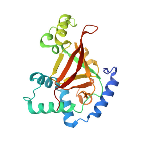 | |
UniProt | |||||
Find proteins for P15879 (Clostridium botulinum D phage) Explore P15879 Go to UniProtKB: P15879 | |||||
Entity Groups | |||||
| Sequence Clusters | 30% Identity50% Identity70% Identity90% Identity95% Identity100% Identity | ||||
| UniProt Group | P15879 | ||||
Sequence AnnotationsExpand | |||||
| |||||
| Ligands 1 Unique | |||||
|---|---|---|---|---|---|
| ID | Chains | Name / Formula / InChI Key | 2D Diagram | 3D Interactions | |
| SO4 Query on SO4 | E [auth B], F [auth C], G [auth D] | SULFATE ION O4 S QAOWNCQODCNURD-UHFFFAOYSA-L |  | ||
| Length ( Å ) | Angle ( ˚ ) |
|---|---|
| a = 105.671 | α = 90 |
| b = 74.951 | β = 102.46 |
| c = 121.199 | γ = 90 |
| Software Name | Purpose |
|---|---|
| REFMAC | refinement |
| DENZO | data reduction |
| SCALEPACK | data scaling |
| AMoRE | phasing |
RCSB PDB (citation) is hosted by
RCSB PDB is a member of the

