2C7W

Crystal Structure of human vascular endothelial growth factor-B: Identification of amino acids important for angiogeninc activity


Experimental Data Snapshot

  • Method: X-RAY DIFFRACTION
  • Resolution: 2.48 Å
  • R-Value Free: 0.310 
  • R-Value Work: 0.286 
  • R-Value Observed: 0.286 

wwPDB Validation   3D Report Full Report


This is version 1.3 of the entry. See complete history


Literature

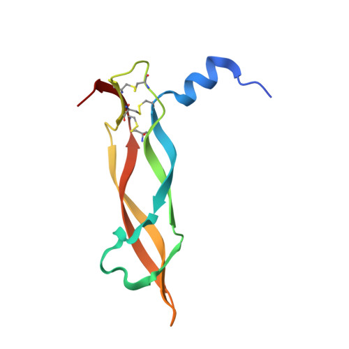
Crystal Structure of Human Vascular Endothelial Growth Factor-B: Identification of Amino Acids Important for Receptor Binding

Iyer, S.Scotney, P.D.Nash, A.D.Acharya, K.R.

(2006) J Mol Biol 359: 76

  • DOI: https://doi.org/10.1016/j.jmb.2006.03.002
  • Primary Citation of Related Structures:  
    2C7W

  • PubMed Abstract: 

    The development of blood vessels (angiogenesis) is critical throughout embryogenesis and in some normal postnatal physiological processes. Pathological angiogenesis has a pivotal role in sustaining tumour growth and chronic inflammation. Vascular endothelial growth factor-B (VEGF-B) is a member of the VEGF family of growth factors that regulate blood vessel and lymphatic angiogenesis. VEGF-B is closely related to VEGF-A and placenta growth factor (PlGF), but unlike VEGF-A, which binds to two receptor tyrosine kinases VEGFR-1 (Flt-1) and VEGFR-2 (Flk-1/KDR), VEGF-B and PlGF bind to VEGFR-1 and not VEGFR-2. There is growing evidence of a role for VEGF-B in physiological and pathological blood vessel angiogenesis. VEGF-B may provide novel therapeutic strategies for the treatment of vascular disease and be a potential therapeutic target in aberrant vessel formation. To help understand at the molecular level the differential receptor binding profile of the VEGF family of growth factors we have determined the crystal structure of human VEGF-B(10-108) at 2.48 Angstroms resolution. The overall structure is very similar to that of the previously determined cysteine-knot motif growth factors: VEGF-A, PlGF and platelet-derived growth factor-B (PDGF-B). We also present a predicted model for the association of VEGF-B with the second domain of its receptor, VEGFR-1. Based on this interaction and the present structural data of the native protein, we have identified several putative residues that could play an important role in receptor recognition and specificity.


  • Organizational Affiliation

    Department of Biology and Biochemistry, University of Bath, Claverton Down, UK.


Macromolecules
Find similar proteins by:  (by identity cutoff)  |  3D Structure
Entity ID: 1
MoleculeChains Sequence LengthOrganismDetailsImage
VASCULAR ENDOTHELIAL GROWTH FACTOR B PRECURSOR
A, B
99Homo sapiensMutation(s): 0 
UniProt & NIH Common Fund Data Resources
Find proteins for P49765 (Homo sapiens)
Explore P49765 
Go to UniProtKB:  P49765
PHAROS:  P49765
GTEx:  ENSG00000173511 
Entity Groups  
Sequence Clusters30% Identity50% Identity70% Identity90% Identity95% Identity100% Identity
UniProt GroupP49765
Sequence Annotations
Expand
  • Reference Sequence
Small Molecules
Ligands 1 Unique
IDChains Name / Formula / InChI Key2D Diagram3D Interactions
MPD
Query on MPD

Download Ideal Coordinates CCD File 
C [auth B](4S)-2-METHYL-2,4-PENTANEDIOL
C6 H14 O2
SVTBMSDMJJWYQN-YFKPBYRVSA-N
Experimental Data & Validation

Experimental Data

  • Method: X-RAY DIFFRACTION
  • Resolution: 2.48 Å
  • R-Value Free: 0.310 
  • R-Value Work: 0.286 
  • R-Value Observed: 0.286 
  • Space Group: P 64
Unit Cell:
Length ( Å )Angle ( ˚ )
a = 120.806α = 90
b = 120.806β = 90
c = 39.826γ = 120
Software Package:
Software NamePurpose
CNSrefinement
HKL-2000data reduction
HKL-2000data scaling
AMoREphasing

Structure Validation

View Full Validation Report


Entry History 

Deposition Data

Revision History  (Full details and data files)

  • Version 1.0: 2007-01-30
    Type: Initial release
  • Version 1.1: 2011-05-08
    Changes: Version format compliance
  • Version 1.2: 2011-07-13
    Changes: Version format compliance
  • Version 1.3: 2023-12-13
    Changes: Data collection, Database references, Other, Refinement description