2C5W

PENICILLIN-BINDING PROTEIN 1A (PBP-1A) ACYL-ENZYME COMPLEX (CEFOTAXIME) FROM STREPTOCOCCUS PNEUMONIAE


Experimental Data Snapshot

  • Method: X-RAY DIFFRACTION
  • Resolution: 2.55 Å
  • R-Value Free: 0.255 
  • R-Value Work: 0.234 
  • R-Value Observed: 0.234 

wwPDB Validation   3D Report Full Report


Ligand Structure Quality Assessment 


This is version 2.1 of the entry. See complete history


Literature

Crystal Structure of Penicillin-Binding Protein 1A (Pbp1A) Reveals a Mutational Hotspot Implicated in Beta-Lactam Resistance in Streptococcus Pneumoniae.

Contreras-Martel, C.Job, V.Di Guilmi, A.-M.Vernet, T.Dideberg, O.Dessen, A.

(2006) J Mol Biol 355: 684

  • DOI: https://doi.org/10.1016/j.jmb.2005.10.030
  • Primary Citation of Related Structures:  
    2C5W, 2C6W

  • PubMed Abstract: 

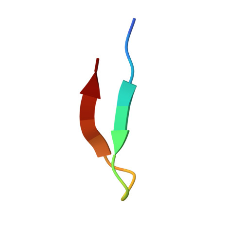
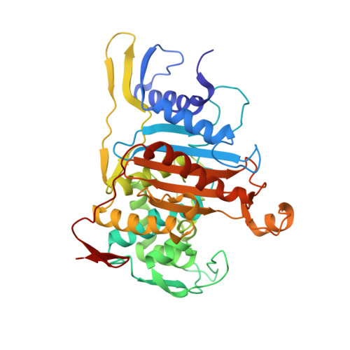
    Streptococcus pneumoniae is a major human pathogen whose infections have been treated with beta-lactam antibiotics for over 60 years, but the proliferation of strains that are highly resistant to such drugs is a problem of worldwide concern. Beta-lactams target penicillin-binding proteins (PBPs), membrane-associated enzymes that play essential roles in the peptidoglycan biosynthetic process. Bifunctional PBPs catalyze both the polymerization of glycan chains (glycosyltransfer) and the cross-linking of adjacent pentapeptides (transpeptidation), while monofunctional enzymes catalyze only the latter reaction. Although S. pneumoniae has six PBPs, only three (PBP1a, PBP2x, PBP2b) are major resistance determinants, with PBP1a being the only bifunctional enzyme. PBP1a plays a key role in septum formation during the cell division cycle and its modification is essential for the development of high-level resistance to penicillins and cephalosporins. The crystal structure of a soluble form of pneumococcal PBP1a (PBP1a*) has been solved to 2.6A and reveals that it folds into three domains. The N terminus contains a peptide from the glycosyltransfer domain bound to an interdomain linker region, followed by a central, transpeptidase domain, and a small C-terminal unit. An analysis of PBP1a sequences from drug-resistant clinical strains in light of the structure reveals the existence of a mutational hotspot at the entrance of the catalytic cleft that leads to the modification of the polarity and accessibility of the mutated PBP1a active site. The presence of this hotspot in all variants sequenced to date is of key relevance for the development of novel antibiotherapies for the treatment of beta-lactam-resistant pneumococcal strains.


  • Organizational Affiliation

    Institut de Biologie Structurale Jean-Pierre Ebel (CNRS/CEA/UJF), Laboratoire de Cristallographie Macromoléculaire, 41 rue Jules Horowitz, Grenoble 38027, France.


Macromolecules

Find similar proteins by:  Sequence   |   3D Structure  

Entity ID: 1
MoleculeChains Sequence LengthOrganismDetailsImage
PENICILLIN-BINDING PROTEIN 1A16Streptococcus pneumoniae R6Mutation(s): 0 
UniProt
Find proteins for Q8DR59 (Streptococcus pneumoniae (strain ATCC BAA-255 / R6))
Explore Q8DR59 
Go to UniProtKB:  Q8DR59
Entity Groups  
Sequence Clusters30% Identity50% Identity70% Identity90% Identity95% Identity100% Identity
UniProt GroupQ8DR59
Sequence Annotations
Expand
  • Reference Sequence
Find similar proteins by:  (by identity cutoff)  |  3D Structure
Entity ID: 2
MoleculeChains Sequence LengthOrganismDetailsImage
PENICILLIN-BINDING PROTEIN 1A385Streptococcus pneumoniae R6Mutation(s): 0 
UniProt
Find proteins for Q8DR59 (Streptococcus pneumoniae (strain ATCC BAA-255 / R6))
Explore Q8DR59 
Go to UniProtKB:  Q8DR59
Entity Groups  
Sequence Clusters30% Identity50% Identity70% Identity90% Identity95% Identity100% Identity
UniProt GroupQ8DR59
Sequence Annotations
Expand
  • Reference Sequence
Small Molecules
Ligands 4 Unique
IDChains Name / Formula / InChI Key2D Diagram3D Interactions
CEF
Query on CEF

Download Ideal Coordinates CCD File 
L [auth B]CEFOTAXIME, C3' cleaved, open, bound form
C14 H15 N5 O5 S2
NRYMPLKBKFIWQC-YVCCLBOHSA-N
ZN
Query on ZN

Download Ideal Coordinates CCD File 
C [auth B],
D [auth B],
E [auth B],
F [auth B],
G [auth B]
ZINC ION
Zn
PTFCDOFLOPIGGS-UHFFFAOYSA-N
EDO
Query on EDO

Download Ideal Coordinates CCD File 
J [auth B],
K [auth B]
1,2-ETHANEDIOL
C2 H6 O2
LYCAIKOWRPUZTN-UHFFFAOYSA-N
CL
Query on CL

Download Ideal Coordinates CCD File 
H [auth B],
I [auth B]
CHLORIDE ION
Cl
VEXZGXHMUGYJMC-UHFFFAOYSA-M
Experimental Data & Validation

Experimental Data

  • Method: X-RAY DIFFRACTION
  • Resolution: 2.55 Å
  • R-Value Free: 0.255 
  • R-Value Work: 0.234 
  • R-Value Observed: 0.234 
  • Space Group: C 2 2 21
Unit Cell:
Length ( Å )Angle ( ˚ )
a = 112.376α = 90
b = 183.373β = 90
c = 54.85γ = 90
Software Package:
Software NamePurpose
XDSdata reduction
XSCALEdata scaling
PHASERphasing
SHARPphasing
CNSrefinement

Structure Validation

View Full Validation Report


Ligand Structure Quality Assessment 


Entry History 

Deposition Data

Revision History  (Full details and data files)

  • Version 1.0: 2005-12-07
    Type: Initial release
  • Version 1.1: 2011-08-03
    Changes: Database references, Derived calculations, Non-polymer description, Other, Refinement description, Structure summary, Version format compliance
  • Version 2.0: 2018-10-03
    Changes: Advisory, Atomic model, Data collection, Derived calculations, Non-polymer description, Structure summary
  • Version 2.1: 2023-12-13
    Changes: Data collection, Database references, Derived calculations, Other, Refinement description