2C57

H.pylori type II dehydroquinase in complex with FA1


Experimental Data Snapshot

  • Method: X-RAY DIFFRACTION
  • Resolution: 3.10 Å
  • R-Value Free: 0.237 
  • R-Value Work: 0.207 
  • R-Value Observed: 0.210 

wwPDB Validation   3D Report Full Report


Ligand Structure Quality Assessment 


This is version 1.3 of the entry. See complete history


Literature

Crystal Structures of Helicobacter Pylori Type II Dehydroquinase Inhibitor Complexes: New Directions for Inhibitor Design.

Robinson, D.A.Stewart, K.A.Price, N.C.Chalk, P.A.Coggins, J.R.Lapthorn, A.J.

(2006) J Med Chem 49: 1282

  • DOI: https://doi.org/10.1021/jm0505361
  • Primary Citation of Related Structures:  
    2C4V, 2C4W, 2C57

  • PubMed Abstract: 

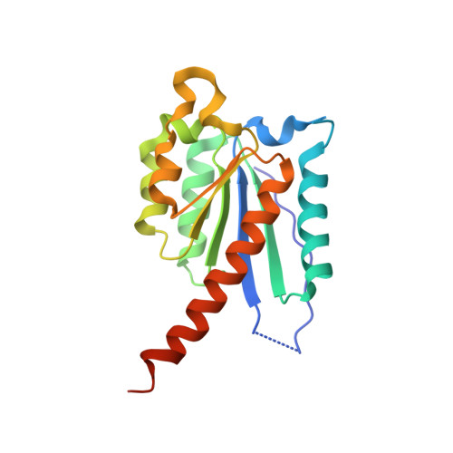
    The crystal structures of the type II dehydroquinase (DHQase) from Helicobacter pylori in complex with three competitive inhibitors have been determined. The inhibitors are the substrate analogue 2,3-anhydroquinate (FA1), citrate, and an oxoxanthene sulfonamide derivative (AH9095). Despite the very different chemical nature of the inhibitors, in each case the primary point of interaction with the enzyme is via the residues that bind the C1 functionalities of the substrate, 3-dehydroquinate, i.e., N76, H102, I103, and H104. The DHQase/AH9095 complex crystal structure shows that sulfonamides can form a scaffold for nonsubstrate-like inhibitors and identifies a large conserved hydrophobic patch at the entrance to the active site as a locus that can be exploited in the development of new ligands.


  • Organizational Affiliation

    Division of Biochemistry and Molecular Biology, Institute of Biomedical and Life Sciences, University of Glasgow, Glasgow G12 8QQ, Scotland, UK.


Macromolecules
Find similar proteins by:  (by identity cutoff)  |  3D Structure
Entity ID: 1
MoleculeChains Sequence LengthOrganismDetailsImage
3-DEHYDROQUINATE DEHYDRATASE
A, B, C, D, E
A, B, C, D, E, F, G, H, I, J, K, L
180Helicobacter pyloriMutation(s): 0 
EC: 4.2.1.10
UniProt
Find proteins for Q48255 (Helicobacter pylori (strain ATCC 700392 / 26695))
Explore Q48255 
Go to UniProtKB:  Q48255
Entity Groups  
Sequence Clusters30% Identity50% Identity70% Identity90% Identity95% Identity100% Identity
UniProt GroupQ48255
Sequence Annotations
Expand
  • Reference Sequence
Small Molecules
Ligands 1 Unique
IDChains Name / Formula / InChI Key2D Diagram3D Interactions
FA1
Query on FA1

Download Ideal Coordinates CCD File 
M [auth A]
N [auth B]
O [auth C]
P [auth D]
Q [auth E]
M [auth A],
N [auth B],
O [auth C],
P [auth D],
Q [auth E],
R [auth F],
S [auth G],
T [auth H],
U [auth I],
V [auth J],
W [auth K],
X [auth L]
2,3 -ANHYDRO-QUINIC ACID
C7 H10 O5
VTEDVYGIJPLVFF-XAHCXIQSSA-N
Binding Affinity Annotations 
IDSourceBinding Affinity
FA1 PDBBind:  2C57 Ki: 3.70e+5 (nM) from 1 assay(s)
BindingDB:  2C57 Kd: 3.70e+5 (nM) from 1 assay(s)
Experimental Data & Validation

Experimental Data

  • Method: X-RAY DIFFRACTION
  • Resolution: 3.10 Å
  • R-Value Free: 0.237 
  • R-Value Work: 0.207 
  • R-Value Observed: 0.210 
  • Space Group: P 31
Unit Cell:
Length ( Å )Angle ( ˚ )
a = 103.863α = 90
b = 103.863β = 90
c = 217.531γ = 120
Software Package:
Software NamePurpose
REFMACrefinement
DENZOdata reduction
SCALEPACKdata scaling
AMoREphasing

Structure Validation

View Full Validation Report


Ligand Structure Quality Assessment 


Entry History 

Deposition Data

Revision History  (Full details and data files)

  • Version 1.0: 2006-02-22
    Type: Initial release
  • Version 1.1: 2011-07-13
    Changes: Advisory, Refinement description, Version format compliance
  • Version 1.2: 2019-05-08
    Changes: Data collection, Experimental preparation, Other
  • Version 1.3: 2023-12-13
    Changes: Data collection, Database references, Other, Refinement description