2C2Q

The crystal structure of mismatch specific uracil-DNA glycosylase (MUG) from Deinococcus radiodurans. Inactive mutant Asp93Ala.


Experimental Data Snapshot

  • Method: X-RAY DIFFRACTION
  • Resolution: 1.70 Å
  • R-Value Free: 0.244 
  • R-Value Work: 0.208 
  • R-Value Observed: 0.209 

wwPDB Validation   3D Report Full Report


This is version 1.3 of the entry. See complete history


Literature

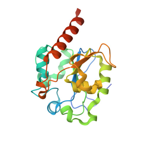
The Crystal Structure of Mismatch Specific Uracil-DNA Glycosylase (Mug) from Deinococcus Radiodurans Reveals a Novel Catalytic Residue and Broad Substrate Specificity

Moe, E.Leiros, I.Smalas, A.O.Mcsweeney, S.

(2006) J Biol Chem 281: 569

  • DOI: https://doi.org/10.1074/jbc.M508032200
  • Primary Citation of Related Structures:  
    2C2P, 2C2Q

  • PubMed Abstract: 

    Deinococcus radiodurans is extremely resistant to the effects of ionizing radiation. The source of the radiation resistance is not known, but an expansion of specific protein families related to stress response and damage control has been observed. DNA repair enzymes are among the expanded protein families in D. radiodurans, and genes encoding five different uracil-DNA glycosylases are identified in the genome. Here we report the three-dimensional structure of the mismatch-specific uracil-DNA glycosylase (MUG) from D. radiodurans (drMUG) to a resolution of 1.75 angstroms. Structural analyses suggest that drMUG possesses a novel catalytic residue, Asp-93. Activity measurements show that drMUG has a modified and broadened substrate specificity compared with Escherichia coli MUG. The importance of Asp-93 for activity was confirmed by structural analysis and abolished activity for the mutant drMUGD93A. Two other microorganisms, Bradyrhizobium japonicum and Rhodopseudomonas palustris, possess genes that encode MUGs with the highest sequence identity to drMUG among all of the bacterial MUGs examined. A phylogenetic analysis indicates that these three MUGs form a new MUG/thymidine-DNA glycosylase subfamily, here called the MUG2 family. We suggest that the novel catalytic residue (Asp-93) has evolved to provide drMUG with broad substrate specificity to increase the DNA repair repertoire of D. radiodurans.


  • Organizational Affiliation

    Norwegian Structural Biology Centre, University of Tromsø, N-9037 Tromsø, Norway.


Macromolecules
Find similar proteins by:  (by identity cutoff)  |  3D Structure
Entity ID: 1
MoleculeChains Sequence LengthOrganismDetailsImage
G/U MISMATCH-SPECIFIC DNA GLYCOSYLASE199Deinococcus radiodurans R1 = ATCC 13939 = DSM 20539Mutation(s): 1 
EC: 3.2.2
UniProt
Find proteins for Q9RWF4 (Deinococcus radiodurans (strain ATCC 13939 / DSM 20539 / JCM 16871 / CCUG 27074 / LMG 4051 / NBRC 15346 / NCIMB 9279 / VKM B-1422 / R1))
Explore Q9RWF4 
Go to UniProtKB:  Q9RWF4
Entity Groups  
Sequence Clusters30% Identity50% Identity70% Identity90% Identity95% Identity100% Identity
UniProt GroupQ9RWF4
Sequence Annotations
Expand
  • Reference Sequence
Small Molecules
Ligands 1 Unique
IDChains Name / Formula / InChI Key2D Diagram3D Interactions
ACT
Query on ACT

Download Ideal Coordinates CCD File 
B [auth A]ACETATE ION
C2 H3 O2
QTBSBXVTEAMEQO-UHFFFAOYSA-M
Experimental Data & Validation

Experimental Data

  • Method: X-RAY DIFFRACTION
  • Resolution: 1.70 Å
  • R-Value Free: 0.244 
  • R-Value Work: 0.208 
  • R-Value Observed: 0.209 
  • Space Group: P 6
Unit Cell:
Length ( Å )Angle ( ˚ )
a = 102.491α = 90
b = 102.491β = 90
c = 37.576γ = 120
Software Package:
Software NamePurpose
REFMACrefinement
MOSFLMdata reduction
SCALAdata scaling
MOLREPphasing

Structure Validation

View Full Validation Report


Entry History 

Deposition Data

Revision History  (Full details and data files)

  • Version 1.0: 2005-10-18
    Type: Initial release
  • Version 1.1: 2011-05-07
    Changes: Version format compliance
  • Version 1.2: 2011-07-13
    Changes: Version format compliance
  • Version 1.3: 2023-12-13
    Changes: Data collection, Database references, Other, Refinement description